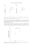
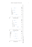
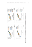
41 Silver Nanoparticles of Bee Honey pH as the skin has an acidic pH of around 4.93 to 5.66. Serum formulations with a pH of about 4.0 to 7.0 are beneficial for the skin as they maintain or even fortify the skin barrier and support the natural skin flora. At the same time, a pH of 4.0 to 5.0 helps reduce the use of preservatives and stabilizes active cosmetic ingredients. The AgNPs–bee honey showed higher activity against gram-positive than gram-negative. In this study, the AgNPs–bee honey serum was prepared and tested against S aureus and E coli. The zone of inhibition in diameter was discerned by disk diffusion method. The clear zone diameter increased as the concentration of AgNPs–bee honey serum increased. The clear zone diameter of the bacterial inhibition zone for S aureus and E coli is shown in Figure 9. The clear zones produced against pathogens tested (1, 2, 3, and 4% of AgNPs–bee honey serum) were found to be 6.1 ± 0.2, 7.9 ± 0.1, 10.2 ± 0.5, 12.3 ± 0.6 mm for S aureus and 0.65 ± 0.02 mm of 4% AgNPs–bee honey serum for E coli, respectively. (a) (b) Figure 9. Zone of inhibition of serum formulations varying the concentration (1–4% AgNPs–bee honey nanoparticles) against S aureus (a) and E coli (b).

42 JOURNAL OF COSMETIC SCIENCE CONCLUSION This study showed that several AgNPs–bee honey nanoparticles had been synthesized successfully. The particle size analysis showed the average particles size was found to be 273 nm with a PDI value of 0.314. The particles showed good antioxidant activity using DPPH radical scavenging assay and FRAP and good antibacterial activity against S aureus. The serum formulation containing 2% bee honey and AgNPs–bee honey nanoparticles was selected due to good stability with preferred homogeneity and pH value, which certainly would make easier the serum application. This work reports a simple, eco-friendly, and economic means of synthesis of AgNPs using bee honey. Future research on its formulation, skin test, and shelf-life performance should be evaluated. ACKNOWLEDGMENTS The author is grateful to the faculty of Science and Technology, Pibulsongkram Rajabhat University, for providing the facilities for working and for the laboratory assistance. REFERENCES (1) P. V. Rao, K. T. Krishnan, N. Salleh, and S. H. Gan, Biological and therapeutic effects of honey produced by honey bees and stingless bees: A comparative review, Braz. J. Pharmacogn., 26, 657–664 (2016). (2) S. H. Ibrahim, N. Z. Soliman, and H. Wissa, Studies on the properties of the major Egyptian honey types and on honey ripening, Agric. Res. Rev., 55, 125–129 (1977). (3) W. N. Julika, A. Ajit, A. Z. Sulaiman, and A. Naila, Physicochemical and microbiological analysis of stingless bees honey collected from local market in Malaysia, Indones. J. Chem., 19, 522–530 (2019). (4) A. B. Jull, N. Cullum, J. C. Dumville, M. J. Westby, S. Deshpande, and N. Walker, Honey as a topical treatment for wounds, Cochrane Database, Syst. Rev., 6, CD005083 (2015). (5) L. S. Chua, N. L. A. Rahaman, N. A. Adnan, and T. T. E. Tan, Antioxidant activity of three honey samples in relation with their biochemical components, J. Anal. Meth. Chem., 313798 (2013). (6) M. K. Rai, S. D. Deshmukh, A. P. Ingle, and A. K. Gade, Silver nanoparticles: The powerful nanoweapon against multidrug-resistant bacteria, J. Appl. Microbiol., 112, 841–852 (2012). (7) F. B. Araruna, P. V. Quelemes, B. E. F. de Faria, S. A. S. Kuckelhaus, V. S. Marangoni, V. Zucolotto, D. A. da Silva, J. R. S. Júnior, J. R. S. A. Leite, and C. Eiras, Green synthesis and characterization of silver nanoparticles reduced and stabilized by cashew tree gum, Adv. Sci. Eng. Med., 5, 890–893 (2013). (8) S. Bogdanov, P. Martin, and C. Lullmann, Harmonised methods of the international honey commission. Liebefeld: Swiss Bee Research Centre, (2002). (9) A. Ali, S. Ambreen, R. Javed, S. Tabassum, I. U. Haq, and M. Zia, ZnO nanostructure fabrication in different solvents transforms physio-chemical, biological and photodegradable properties, Mater. Sci. Eng. C, 74, 137–145 (2017). (10) I. F. F. Benzie and J. J. Strain, The ferric reducing ability of plasma (FRAP) as a measure of antioxidant power: The FRAP assay. Anal. Biochem., 239, 70–76 (1996). (11) P. V. Quelemes, F. B. Araruna, B. E. F. De Faria, S. A. S. Kuckelhaus, D. A. da Silva, R. Z. Mendonça, C. Eiras, M. J. S. Soares, and J. R. S. A. Leite, Development and antibacterial activity of cashew gum based silver nanoparticles, Int. J. Mol. Sci., 14, 4969–4981 (2013). (12) P. Singh, Y. J. Kim, H. Singh, C. Wang, K. H. Hwang, M. E. A. Farh, and D. C. Yang, Biosynthesis, characterization and antimicrobial applications of silver nanoparticles, Int. J. Nanomedicine, 10, 2567– 2577 (2015).
Purchased for the exclusive use of nofirst nolast (unknown)
From: SCC Media Library & Resource Center (library.scconline.org)