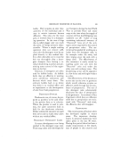
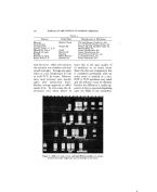
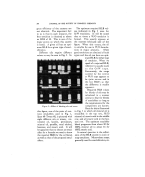
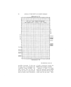
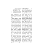
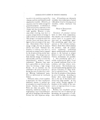
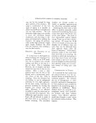
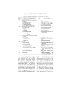
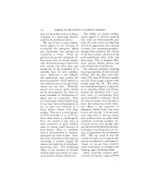
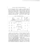
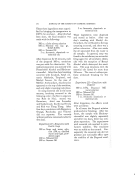
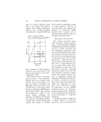
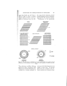
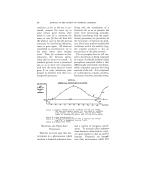
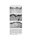
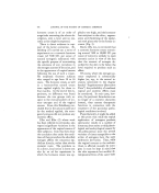
410 JOURNAL OF THE SOCIETY OF COSMETIC CHEMISTS Figure 2.--Absence of response in skin of a young woman. •/, Biopsy specimen from the skin of the back before treatment. Note the essentially normal epidermis with large, well-developed pegs and vesicular cells B, biopsy specimen taken after inunction of estrogen- free ointment for sixty days. There is no perceptible change C, biopsy specimen taken after inunction of ointment containing 242 international units of estrogen per gram. There is no difference between this and the control biopsy specimen. (Reproduced from the paper by Eller and Eller in the Airchives of Dermatology and Syphilology, 59, 449 (1949).)

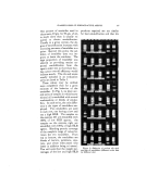
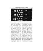
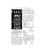
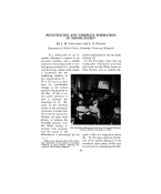
COSMETIC ASPECTS OF ESTROGENIC HORMONES 411 porary character of this procedure by stressing the decline of its effec- tiveness upon the discontinuance of the application of the hormone ::. cream. An extens)ve investigation of hormone creams was carried out by Eller and Eller ($). It was found i.i' that estrogenic hormone ointments (with 7500 to 15,000 I.U. per ounce of excipient) produced consistent :.: proliferative effects upon the skin epithelium (Fig. 1), the response varying with the age of the subject, the concentration of the estrogen, the duration of the individual ap- ß plication, and the total duration of the treatment no epithelial response was obtained in the case of young women (20 to 30 years of age) who were free of any symptoms of endocrine dysfunction and who exhibited a fully developed epidermis of normal thickness (Fig. 2). Definite histological changes were observed in all 19 post-menopausal subjects (aged 51 to 79) whose con- trol biopsies showed senile atrophy with thinning of the epithelium. These changes were: (a) the cytoplasm tended to in- crease in volume, (b) the nuclei increased in size and assumed a rounded rather than ovoid form, (c) the basal cell layer increased in wariness indicating proliferative activity, •' (d) the epidermis gradually in- creased in thickness, and the epider- mal pegs reappeared, (e) the capillary blood supply became more prominent, (f) the supportive elastic fibrillar structures became more pronounced, (g) the elastic tissue and colla- gen (of the underlying corium) was affected only to a minimal degree. The changes in the skin's epithe- lium reached a maximum at some time between thirty and fifty days this maximum was attained more promptly with the large doses, and with the more prolonged periods of contact with tke estrogenic material. They were maintained as long as the application was continued omis- sion of treatment caused reversion to the original atrophic condition. There was a latent period of ten to twenty days before the appearance of a demonstrable effect. SUPPLEMENTARY ILLUSTRATIVE DATA j. Goldzieher (6) studied the effect upon tke senile skin of estrogenic (and androgenic) hormones applied by spraying (in the form of alcohol- ether solutions). While biopsies re- vealed distinct regenerative changes in the epidermis, his findings are of a lesser relevance to the consideration of the subject matter of this presen- tation, since the concentrations of the steroids employed were far above the physiologic range. By way of supplementary in- formation, reference is made to sev- eral selected papers which bear upon the general problem of local action of estrogenic hormones. It has been known for some time that topical hormone therapy gave ex- cellent results in kraurosis vulvae and other regressive conditions of the
Purchased for the exclusive use of nofirst nolast (unknown)
From: SCC Media Library & Resource Center (library.scconline.org)