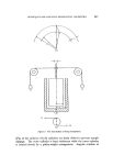
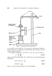
THIN-LAYER CHROMATOGRAPHIC TECHNIQUES IN RESIDUE ANALYSIS 485 very good contrast and definition. Either positive or negative copies can be obtained on these papers and the process is equally suitable for copying paper chromatograms, graphs, typescripts, etc., although longer exposures are required. To copy a chromatoplate, either circumscribe the spots with a soft-lead pencil or scrape them completely from the plate. Similarly mark the origin points and the solvent front any other information may also be inscribed on the layer, writing letters backwards and from right to left. Remove excised material by gently blowing and place the marked plate face downward on the sensitized surface of a sheet of the diazo-paper 10 x 8" is a convenient size when using 20 x 20 cm chromatoplates. Illuminate from above with four 275w photoflood lamps mounted in a reflector (25 x 25 x 20 cm) placed centrally over the plate. After exposing for from 5 to 15 sec, depending on the speed of the paper, remove the light source and suspend the paper in an atmosphere of ammonia. The marked spots appear almost immediately as white rings or spots on a coloured back- ground. The copies so obtained are permanent, readily filed for later inspection and may be inscribed as desired. Quantitative evaluation of chromatograms The techniques which have been applied to the quantitative evaluation of thin-layer chromatograms fall readily into two classes. That which has the largest following among pesticide residue analysts comprises those pro- cedures in which the chromatographed compound is separated from the layer absorbent before the application of standard micro-chemical analytical methods such as spectrophotometry, gas-liquid chromatography, etc. The alternate technique involves the relationships which exist between the weight of compound contained in the located spot and its size, density of coloration (visual or photometric), or radioactivity. The very small amounts (micrograms) of material usually applied to a thin-layer plate precludes the use of simple weighing of the eluted com- pound unless many chromatoplates are developed and the products combined (100). Spectrophotometry in the visible and uv regions of the spectrum can readily be carried out with the weights of material (0-10 •g) which may be eluted from a single spot on a chromatoplate. Kirchner et al (9) used uv spectrophotometry to determine diphenyl eluted from silica gel Deters (101) has similarly determined traces of pentachloro- phenol. With these techniques some interference may be observed from extraneous material contained in the absorbent layer. Pre-extraction with

486 JOURNAL OF THE SOCIETY OF COSMETIC CHEMISTS methanol has been found (102) to reduce this interference, but the pre- paration of a blank extract of an adjacent area of the layer was still necessary for full correction other errors were introduced by incomplete elution from the absorbent. Dimethoate and its oxygen analogue have been determined in vegetable tissue by Steller and Curry (81) who digested the eluted pesticides with a nitric-perchloric acid mixture and evaluated the liberated phosphorus by a molybdenum blue spectrophotometric procedure. Dinoseb has been determined spectrophotometrically as its sodium salt after a clean-up on wedge-layer plates (36). Infra-red spectrometry has also been used for the quantitative deter- mination of organo-phosphorus pesticides (34) as well as simultaneously providing conclusive evidence of the identity of the separated pesticide. Gas-liquid chromatography provides a very convenient method for the determination and further clean-up of the eluted pesticide spots. The use of electron-capture detection for both organo-chlorine and organo- phosphorus compounds (41,103) gives very great sensitivity. The use of a silver nitrate-uv irradiation procedure for the visualization of the chromatogram leaves the bulk of the pesticide unaffected in each spot since only the surface of the layer comes under the influence of the irradiation. Thus elution of the pesticide from the treated chromato- plate with dichloromethane gives ample material for an approximately quantitative evaluation. If insufficient material is present to give a visible spot with the visualization reagent used, then elution of that area of the layer suspected of containing the pesticide may give sufficient material for further examination by gas-liquid chromatography. Biological assay, using Drosophila melanogaster, has been used by Salo et al (79) to determine some organo-chlorine and organo-phosphorus pesticides separated on silica gel containing built-in fluorescein as indicator. Marco and Jaworski (104) utilized C •4- labelled Colep for the determination of residues of the pesticide and its various metabolites by radiometry. A suitable scanner for thin-layer chromatography has been described by Wilde (105). A useful description of the sequence of techniques involved in the routine quantitative analysis of materials which have been separated by thin-layer chromatography has been given by Millett, Moore and Seaman (106). Application of the samples, removal of the layer material bearing the located compound, and the elution of the latter from the absorbent are detailed the accuracy and precision (1-2%) of the techniques are also discussed.
Purchased for the exclusive use of nofirst nolast (unknown)
From: SCC Media Library & Resource Center (library.scconline.org)