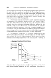
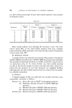
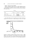
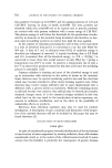
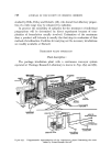
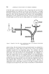
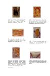
746 JOURNAL OF THE SOCIETY OF COSMETIC CHEMISTS hair growth ceased. However after a short lag period derreal papillae of apparently normal size were regenerated and whiskers of normal length were grown. Even when the complete bulb region and up to the lower third of the follicle were removed, smaller than normal dermal papillae regener- ated and generations of short whiskers were produced. These papillae regenerated at the level of tissue removal and were not accompanied by a lengthening of the follicle. It was further found that the greater the length of follicle removed the shorter were the whiskers subsequently grown. Although papilla regeneration and whisker growth did not occur where more than the lower third of the follicle was removed, the remaining part persisted. The mesenchymal layer, the outer root sheath, and their glassy mem- brane interface were identified as the essential prerequisites for this re- generation phenomenon since segments of the follicle wall, provided that their proximal ends derived from within the lower third of the follicle, regenerated papillae and produced whiskers when implanted into ear dermis (36). The new papillae apparently developed from cells from the mesenchymal layer. The preservation of the tubular nature of the implants was also shown to be important because when they were incised longitudinally, opened out and implanted as sheets they were unable to organize follicles and degenerated (37). The derreal papilla and the induction of hair growth Failure of papilla regeneration in the upper two thirds of the follicle, while in itself an intriguing finding, nevertheless enabled the strongly suspected inductive property of the dermal papilla to be tested. Dermal papillae, completely free of contaminating epidermal matrix cells, were implanted into the bases of the superficial halves of follicles after removal of their lower halves (38). Where the papillae remained in position the growth of generations of shorter than normal whiskers ensued. An important difference was observed in these follicles when compared with those which regenerated papillae after tissue removal. Associated with the induction of bulb formation and whisker growth the follicle grew in length as it does during anagen in the normal pelage hair cycle (Fig. 2). Cells from the implanted papilla were also seen to contribute to the mesen- chymal layer over the newly grown length of follicle. As a logical extension of these results the influence of whisker dermal papillae on epidermis of non-whisker origin was studied.

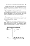
DERMAL PAPILLA AND THE DEVELOPMENT AND GROWTH OF HAIR 747 The dermal papilla and its influence on ectopic epidermis in ear dermis Cohen (31, 30) and Oliver (35) have shown that the complete whisker bulb region and also papillae with just their own epidermal investments can generate follicles which produce short whiskers when implanted into ear dermis, although the follicles did not develop typical whisker dermal features. Cohen (31, 30, 40) has also implanted whisker dermal papillae alone into ear dermis. He considered that where they contacted ear epidermis they induced the development of ear hair follicles within 14 days, the number of papilla cells being reduced from hundreds down to tens to comply with the normal ear follicle configuration. Adopting a slightly different approach Oliver (41, 42) has made an extended study of the influence of whisker dermal papillae on ectopic epidermis in ear dermis and the collective results will now be considered. Two types of surface epidermis were used, ear epidermis containing follicular epidermal elements and afollicular scrotal sac epidermis, and keratinizing oral epithelium from either the gum or inner lip regions. Small sheets of epidermis or epithelium were obtained by trypsinization and placed basal surface down onto groups of two to six papillae on an ear dermis bed. The implants were then covered by replacing a previously reflected flap of skin and the graft areas biopsied from 7-207 days after operation and serially sectioned. The papillae did not always remain in contact with the grafted epidermis or epithelium and were found isolated in ear dermis, or in contact with ear epidermis at the edge of the implant region, as well as associated with the graft epidermis or epithelium. Approximately 370//0 of the implanted dermal papillae were found isolated in ear dermis. All of these papillae were entirely free of adherent epidermal cells, corroborating the observed absence of such epidermal cells by examination of serial sections of papillae dissected in exactly the same manner ($8). In general they seemed to have maintained approximately their original number of cells, although they were often compacted with a decrease in cellular cytoplasmic volume. Most had acquired a capillary supply and stained with alcian blue. Obviously their integrity was not dependent on contact with epidermal cells. Eighteen dermal papilla/ear epidermis associations were examined. From days 7-21 large alcian blue staining papillae with capillaries had locally induced epidermal hyperplasia, with the epidermis becoming organized as matrices, with suprabasal mitoses, around the papillae. In the single day, 28 specimen vacuolated outer root sheath and an inner root
Purchased for the exclusive use of nofirst nolast (unknown)
From: SCC Media Library & Resource Center (library.scconline.org)