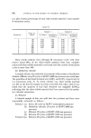
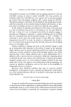
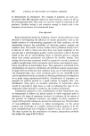
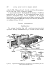
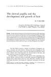
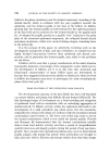
748 JOURNAL OF THE SOCIETY OF COSMETIC CHEMISTS sheath containing characteristic trichohyalin granules had also developed, but no other follicular features were present. Between days 35-64, eight follicles induced by single papillae and all producing hair were examined. All were shorter, stouter and had larger bulbs than metanagen ear hair follicles and their papillae contained hundreds of cells compared with the approximately 30-50 present in ear hair follicles (Fig. $). Nevertheless the number of cells incorporated into the papillae of the induced follicles was less than the number present at implantation, accountable in part by the absence of capillaries which are a feature of normal whisker papillae but not of ear hair papillae. Five of the follicles were growing fine non-medullated hairs associated with a thick inner root sheath, while three had thick whisker-like shafts, one at least of which was medullated. Seven of the follicles contained differentiating sebaceous glands but, with the exception of one which opened on to the ear surface, had no sebaceous ducts. The inner root sheath was present at least as far as the ear surface in all of the follicles {Fig. •t), which also showed varying degrees of dermal sheath differentiation but without the development of whisker follicle features. An exception to the developmental sequence outlined above was the presence, at day 63, of a single papilla associated with a matrix at the ear surface which was producing thick laminate keratin. Distinct trichohyalin granules were present at the periphery of the matrix as were some sebaceous cells. With afollicular scrotal sac epidermis whisker dermal papillae were also seen to organize matrices and at day 35 two very large intradermal follicular structures were found, with the usual epidermal elements except sebaceous cells, which were producing hair keratin. Implants of oral epithelium and its own dermis or oral epithelium alone, regardless of whether they formed cysts or became incorporated into the ear surface, showed the preservation of the typically oral pattern of kera- tinization, with the absence of a granular layer and of appendages. This accords with the findings of Billingham and Silvers (23) with tongue and oesophageal epithelium, recombined with ear and sole of foot dermis, in the guinea pig and hamster. In marked contrast a picture of confusion of influence and response was shown by the 22 examples of dermal papilla/oral epithelium associations examined between 24 and 64 days. Contiguous papillary tissue promoted epithelial hyperplasia, with supra- basal mitoses, and epithelial thickening, the basal cells often being highly columnar {Fig. $). Two types of "projections" were seen, one being large

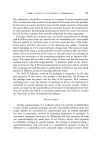
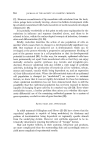
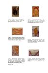
DERMAL PAPILLA AND THE DEVELOPMENT AND GROWTH OF HAIR 749 with a central core of papillary tissue containing capillaries. Here the inner aspect of the epithelium was ridged and thick squamous keratin was being produced. Alternatively, associated with smaller papillary indentations, there was the localized development of large fibrous cells, resembling cortex cells in the keratogenous zone of hair follicles, which were producing spikes of keratin, as opposed to squames, histologically identical to the alpha- keratin of hair. There were also instances of epithelium present as a stellate reticulum reminiscent of the enamel organ of developing teeth (Fig. $). Several small hair folhcles were found as were, in other regions, sebaceous cells sometimes associated with the locahzed development of a granular layer. Otherwise a large, very short, bulb-like configuration was seen with lateral outer and inner root sheaths whose matrix was producing large, loosely associated cortex-like cells containing wispy keratin fibres (Fig. 6). There was also a large follicular structure with follicle sheaths but, instead of differentiating hair elements, the papilla was capped by a stellate reti- culum configuration. Finally, incorporated into one side of a papilla/epi- thelial complex, was a small aberrant follicle with the usual follicular features on the ear dermis side, which was producing a spike of keratin via large fibrous cells as described for the small type of projection above. DISCUSSION The derreal papilla and follicle development The prospective dermal papilla is established as a permanent and stable population of cells very early in the ontogenetic development of the hair follicle, appearing as an aggregation of cells at or before the first histological signs of change in the epidermis. There is evidence that a crucial interaction occurs at this time fixing the site of follicle development (15). After this interaction occurs the epidermal component presumably also has an active developmental role since in certain circumstances it can initiate the develop- ment of new papillae (17, 18, 23). As follicle development proceeds a second vital interactive stage is reached which is essential to ensure hair growth (15). The factors responsible for establishing dermal papillae as specialized variants of the dermis are unknown. Presumably they arise as a consequence of other sub- and intradermal events related, perhaps, to the timing and pattern of development of the peripheral neural and vascular systems (43, 44). Whatever the reason, it is known that follicles can develop in vitro in embryonic skin explanted days before follicle histogenesis such explants
Purchased for the exclusive use of nofirst nolast (unknown)
From: SCC Media Library & Resource Center (library.scconline.org)