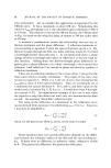
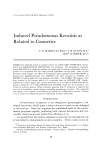
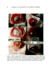
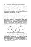
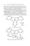
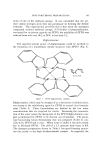
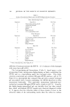
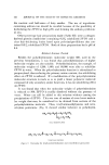
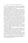
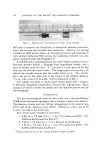
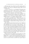
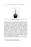
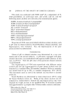
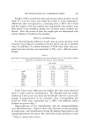
PSEUDOMONAS KERATITIS 91 into the cornea. When the cornea* was cut sharply with a scalpel partway into the stroma, the subsequent instillation of P. aeruginosa into the con- junctival sac routinely resulted in pseudomonas keratitis. It thus became apparent that the epithelium constituted an important protective barrier damage had to extend into the stroma to produce infection. Definitive Studies Experiments on Corneas This test was designed to determine the number of pseudomonas or- ganisms required to produce pseudomonas keratitis in the rabbit when instilled into the conjunctival sac, the time-course of events in the devel- opment of pseudomonas keratitis, and the toxicity of pseudomonas endo- toxin. Both eyes of each of 16 adult New Zealand White rabbits were admin- istered three parallel cuts with a scalpel through the epithelium and slightly into the stroma. Less than 1 min later, 0.05 ml from suspensions of P. aeruginosa to yield inocula ranging from 75 to 7.5 million cells (in increments of one order of magnitude) was instilled into each of 24 eyes. A control group of 4 eyes received no organisms and one group of 4 eyes received 0.1 ml of pseudomonas lipopolysaccharide, an endotoxin (7) purified by the method of Webster et al. (8). Results, summarized in Table I, show that as few as 75 organisms of this strain may in some cases produce significant corneal effects in rabbits. The number of organisms of this strain required to produce a positive response in 50% of the animals (EN5o) under these experimental condi- tions appea. rs to lie between 75 and 750. At 24 hours, the affected eyes displayed various degrees of corneal opacity, iritis, chemosis, injection of the lids, and white discharge. Al- though the aqueous humor appeared to be clear, microscopic examina- tion disclosed the presence of an abundance of leucocytes (hypopyon). By 48 hours, the lids became beefy red. Opacity in most cases involved the entire cornea. By 72 hours, all affected eyes showed perilimbal vas- cularization. Perforation of the cornea was observed in two animals in 13 days (Fig. 1). * In these experiments the cornea was anesthetized with Ophthaine© (Proparacaine HC1 ophthalmic soln. 0.5%, E. R. Squibb g: Sons, 909 Third Ave., N.Y., N.Y.) However, results with general anesthesia were found to produce the same effects.

92 JOURNAL OF THE bOCIETY OF COSMETIC CHEMISTS Figure 1. Ocular damage from P. aeruginosa and pseudomonas endotoxin. Rabbit eyes: corneal effects after instillation ,of P. aeruginosa into conjunctival sacs of animals with incised corneas, at 24 hr (top left), at 48 hr (center left), and at 16 days (bottom left) intrasderal effects at 48 hr (top right) effects of intracomeal injection of endotoxin at 9 days (center right). Monkey c)c: 7 da)s after intracorncal injection of P. aerugino.•'a (bottom right)
Purchased for the exclusive use of nofirst nolast (unknown)
From: SCC Media Library & Resource Center (library.scconline.org)