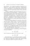
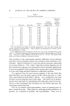
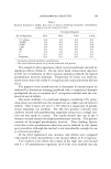
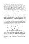
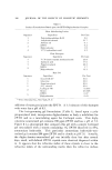
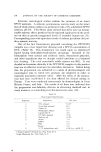
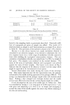
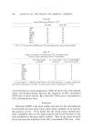
PSEUDOMONAS KERATITIS 93 Table ! Keratitis Produced in Rabbit Eyes (Cut to Stroma) Following Instillation of Pseudomonas aeruginosa into the Conjunctival Sacs Fractional Response No. of Organisms 24 hr 48 hr 72 hr a 6 days 7.5 million 4/4 4/4 4/4 4/4 750,000 4/4 4/4 4/4 4/4 75,000 3/4 4/4 4/4 4/4• 7,500 4/4 4/4 4/4 4/4• 750 3/4 4/4 4/4 4/4 75 1/4 1/4 1/4 1/4 0 0/4 0/4 0/4 0/4 Endotoxin 0,/4 0,/4 0,/4 0,/4 a All positives showed perilimbal vascularization. • One animal showed opacity on •/• cornea, associated with pannus. The animals in ,this experiment which received endotoxin showed no significant effects (Table I). On the other hand, intracorneal injection of 0.01 ml of endotoxin in three separate animals produced the typical pseudomonas keratitis syndrome. Progression of events was, however, much slower than with viable P. aeruginosa cells, and perforation did not Occur. The purpose of the second test was to determine if corneal injury (as indicated by fluorescein staining) produced with a com•nercial sha•npoo predisposed the eye to invasion by P. aeruginosa instilled into the con- junctival sacs of rabbits. One-tenth •nilliliter of a •narketed shampoo containing 13% potas-- sium oleate was instilled into the conjunctival sacs (right eyes) of each of 4 rabbits. After 4 hours, 0.1 ml (7 X 10 4 cells) of a suspension of pseudo- monas organis•ns was instilled. The left eyes (positive controls) were si•nilarly treated with pseudo•nonas organisms except that a scalpel inci- sion was first •nade in corneas. The results showed that one of •the 4 shampoo-treated ani•nals developed pseudo•nonas keratitis. The positive controls all developed pseudmnonas keratitis. These findings demon- strate that ocular pseudo•nonas infections can be induced by this (sha•n- poo) procedure, al,though the •nethod is not reproducible enough for use as a research procedure. In the third exploratory test, monkeys and rabbits were compared with regard to their susceptibility to the invasive effects of P. aeruginosa. Two monkeys were tested ,the corneas of the right eyes were incised and 6 X 10 • pseudomonas organisms (in 0.4 ml) were instilled into the

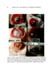
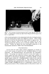
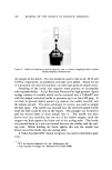
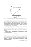
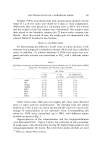
94 JOURNAL OF THE SOCIETY OF COSMETIC CHEMISTS conjunctival sacs the left eyes were injected intracorneally with 0.01 ml (7 X 10⁴ cells) of a suspension of pseudomonas organisms. Four pseudo- monas-treated incised rabbit eyes were used as positive controls. Re- sults of this ,test, though exploratory, strongly suggest that monkey eyes are not as seriously affected by these pseudomonas organisms as are rabbit eyes. All 4 rabbit eyes showed typical pseudomonas keratitis, ending in perforation in 10 days. All 4 monkey eyes showed effects which were less severe than those in rabbit eyes. The maximum effects (in all monkey eyes) occurred in 48-72 hours and consisted of a white corneal opaci. ty with redness of the lids. This persisted for several weeks with gradual improvement but incomplete resolution (Fig. 1). Experiments on Tissues Other Than the Cornea A series of experiments was conducted to investigate •the susceptibility of tissues other than the cornea to invasion from pseudomonas organisms or injury from its products. Both tissues investigated, the sclera and the skin, are vascular tissues, in contrast to the cornea. In addition, their collagens may be different biochemically. Studies on Sclera--Incisions about 5 mm long were made with a scalpel deep into the scleral fibers of three rabbits. Immediately thereafter, 0.1 ml (7.5 million cells) of a suspension of pseudomonas organisms was in- stilled into the conjunctival sacs. Findings were essentially negative. Only slight injection of blood vessels was observed at the incision sites. Intrascleral injection of pseudomonas organisms, however, produced more dramatic effects than those seen above. The right eyes of each of 4 rabbits were injected with 0.02 ml (3 X 10⁵ cells) of a suspension contain- ing pseudomonas organisms. The left eyes (positive con•trols) were ad- ministered about 1.5 X 10 ø cells in the conjunctival sacs after the corneas were incised with a scalpel as described previously. Intrascleral adminis- tration produced upper lid redness and chemosis and a central abscess surrounded by injected blood vessels. Peak effects were reached in 1-3 days with gradual resolution in about 2-3 weeks (Fig. 1). The posi,tive controls all developed'typical pseudomonas keratitis. Studies on Skin--A straight incision about 5 cm long was made with a scalpel deep into the dermis on the clipped backs of 12 animals. Each cut was filled with a suspension containing about 7.5 million pseudo- toohas organisms. Examination of these sites at various times thereafter showed no significant skin reactions. Intradermal injection (0.1 ml/site) of the backs of 2 rabbits with a suspension containing 15 million pseudornonas •cells per site resulted in
Purchased for the exclusive use of nofirst nolast (unknown)
From: SCC Media Library & Resource Center (library.scconline.org)