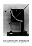
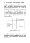
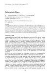
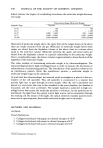
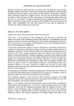
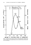
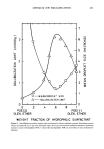
448 JOURNAL OF THE SOCIETY OF COSMETIC CHEMISTS linked dextran the degree of crosslinking determines the molecular weight fractiona- tion range. Fractionation Range (Molecular Weight) Sephadex Type Peptides Dextrans G-10 - 700 - 700 G-15 - 1,500 - 1,500 G-25 1,000- 5,000 100- 5,000 G-75 3,000- 70,000 1,000- 50,000 G-200 5,000-800,000 1,000-200,000 Molecules of molecular weight above the upper limit of the ranges shown in the above chart are totally excluded from the gel. Molecules of molecular weight below these ranges are eluted from the Sephadex column in the above chart at a volume about equal to the total bed volume. Molecules between the upper and lower limits are eluted from the Sephadex column in a specific relationship to the molecular weight. Over a considerable range, the elution volume is approximately a linear function of the logarithm of the molecular weight. The other method of determining molecular weight is by ultracentrifugation. The ultracentrifuge produces high centrifugal forces in order to measure the movement or redistribution of sedimenting particles. The distribution of the particles is observed by an interference pattern. From this interference pattern, a molecular weight or molecular weight range can be calculated. In work with the ultracentrifuge, the material under investigation is placed in the cen- terpiece of the cell assembly. This cell assembly is constructed in a manner that permits light rays to pass through its entire length. After the cell is assembled and placed in a rotor hole, the rotor is then installed in the rotor chamber the chamber is evacuated and the rotor accelerates. The sample material is subjected to high cen- trifugal forces that causes the molecular particles to sediment. As the particles are re- distributed, the light from the optical system light source can be transmitted through the rotating cell. By means of this light, the optical elements translate particle move- ment into an optical pattern, from which molecular weight can be calculated. METHODS AND MATERIAL MATERIALS Protein Hydrolysates 1. Collagen hydrolyzed with papain to a formol nitrogen of 10.0* 2. Collagen hydrolyzed with steam to a formol nitrogen of 6.0* 3. Collagen hydrolyzed with acid to a formol nitrogen of 10.0' *Inolex Corporation, Chicago, IL.

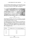
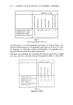
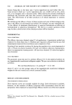
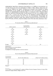
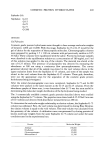
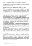
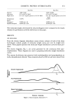
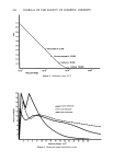
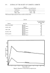
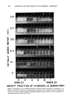
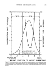
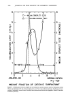
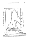
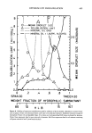
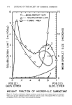
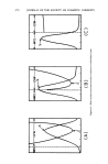
COSMETIC PROTEIN HYDROLYSATES 449 Sephadex Gels Sephadex G-10 G-15 G-25 G-75 G-200 METHODS Gel Filtration Cosmetic grade protein hydrolysates were thought to have average molecular weights of between 1,000 and 10,000. With this range, Sephadex G-15 or G-25 would be the gel of choice for the separation of these peptide molecules. Chromatographic columns were prepared by packing 1.5 x 100 cm columns with gel previously swollen in 0.25 M NaC1. These columns were equilibrated with the saline. Protein hydrolysate samples were dissolved in the equilibration solution at a concentration of 180 mg/ml, and 3 ml of this solution was applied to the top of the column. The material was eluted at the rate of 0.25 ml/min. The presence of polypeptides was detected by measuring the absorbance at 280 nm using a continuous flow spectrophotometer. The curves generated showed that all of the sample was eluted in the void volume (above in the upper exclusion limit) from the Sephadex G-15 column, and some of the sample was eluted in the void volume from the Sephadex G-25 column. These gels, therefore, were not the appropriate ones for the separation of the cosmetic grade protein hydrolysates being investigated. After the initial chromatographic runs were completed, Sephadex G-75 and G-200 columns were packed in the same manner as the G-15 and G-25 columns. From the absorbance graphs of these runs, it was determined that G-75 was the most useful in determining the molecular weight distribution of the hydrolysates being studied. The 3 commercially available cosmetic grade proteins described above were studied using the Sephadex G-75 column. The peptides were eluted with 0.25 M NaC1 at a rate of 0.15 ml/min and detected with absorbance measurements at 280 nm. To determine the molecular weight relationship to elution volume, the Sephadex G-75 column was calibrated. First, the void volume was determined by eluting Blue Dextran the elution volume is equal to the void volume. The total volume was calculated from the geometry of the column. A calibration curve was generated by eluting proteins of known molecular weights from the same Sephadex G-75 column and under the same conditions used in the experimental run. Protein Molecular Weight Aldolase 158,000 Ovalbumin 45,000 Chymotrypsinoge n 25,000 Ribonuclease A 13,700
Purchased for the exclusive use of nofirst nolast (unknown)
From: SCC Media Library & Resource Center (library.scconline.org)