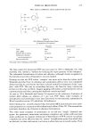
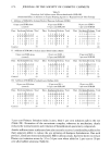
684 JOURNAL OF THE SOCIETY OF COSMETIC CHEMISTS treatment etch solution were analyzed. The difference between the fluoride content of the treated tooth surface and that of the untreated tooth surface gave the amount of fluoride incorporated into the enamel. ANIMAL MODEL SYSTEM FOR ASSESSING CARIES REDUCTION BY FLUORIDE DENTIFRICES The animal assay system employed was essentially that ofK6niget al. (6). It gave an ex- cellent dose-response curve with aqueous fluoride solutions containing fluoride at levels occurring in dentifrices. (The method will only be outlined here details will be reported elsewhere.) Osborne-Mendel albino rats were employed. They were main- tained under conditions generally observed for specific pathogen-free animals. Females were fed a balanced vitamin-supplemented diet from mating to the end of the suckling period. Trials were started on the day of weaning. Weanlings were randomly dis- tributed to the various treatments, the animals being distributed among the cages so as to equalize the stresses of weaning, treatment, and cariogenic diet. A cariogenic diet was fed ad/ibitum. It consisted of sucrose (56 per cent) plus milk powder and other essential nutrients. About 24 animals were subjected to each treatment. Twenty- second applications of the materials to be tested were applied to the lower jaws by means of a marten-hair brush, using about 15 to- and fro- motions of the brush. The rats were deprived of food and water for 1 h after treatment. The treatments were ap- plied twice daily during the first 2 weeks and once daily during the third week (no treatment on Sundays). The rats were sacrificed, and the lower jaws were removed and prepared for sectioning and evaluation of carious lesions after staining. The severity of carious lesions in the first and second molar teeth was assessed by the method of K6nig eta/., which grades carious lesions in terms of the stages of the carious process from start (in the enamel) through the next stage (in the dentin) to the conclusion (cavita- tion). In essence, 4 stages are recognized after staining: A lesions (limited to enamel, no staining of the adamantine border), T lesions (early dentinal lesion, involving color reaction at the adamantine border), B lesions (moderate dentin lesion, comprising progression of the lesion with decalcificiation of dentin bordering on the pulp), and C lesions (severe dentin lesion, involving destruction in the direction of both the pulp and occlusal surfaces, loss of enamel in the sulcus, and first signs of cavitation). In calcu- lating the reduction of incidence of carious lesions produced by the test products com- pared to nonfluoride products in the current study, only the more severe lesions (B and C) were considered. These values were combined, and reduction in carious lesions cal- culated as: Per cent reduction lesions = 100 x (B + C lesions on nonfluoride dentifrice) - (B + C lesions on fluoride dentifrice) (B + C lesions on nonfluoride dentifrice) Of course, the values obtained by this procedure are valid for only within-trial com- parisons. Also, it should be recognized that the magnitude of the reduction in nu- merical terms can depend on the value attributed to the various lesions in this study it was found advantageous to base the calculatons of fluoride effect in the rat on only the B and C lesions.

Purchased for the exclusive use of nofirst nolast (unknown)
From: SCC Media Library & Resource Center (library.scconline.org)