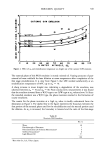
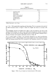
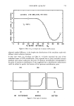
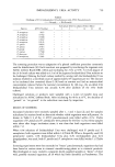
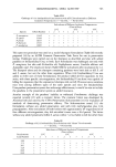
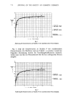
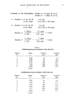
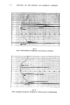
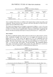
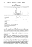
780 JOURNAL OF THE SOCIETY OF COSMETIC CHEMISTS Middleton worked with guinea pig foot pads and found a 28.2% increase over dry weight at 90% R.H. at room temperature (4). Fox et al. worked with pulverized, un- washed human callus and found an 11% increase over dry weight at 60% R.H. and 34% increase at 90% R.H. at 23øC (15). These values are similar to our cantharidin data. For comparison, Table 4 presents values that other workers have reported with various methods. One might draw the conclusion that callus and stratum corneum obtained from the back by cantharidin are analogous in their physical chemical properties however, Blank and Shappirio pointed out that water extraction of callus for 2 hr did not alter water holding capacity, but did decrease flexibility (16). Different stratum corneum harvest methods may lead to differences in hygroscopicity, but the differences cannot be demonstrated to be statistically significant. Yet, a Table IV Comparison of Stratum Corneum Hygroscopicity % Increase % Relative Over Investigator Source of Corneum Temp. øC Humidity Dry Weight Singer & Vinson (10) Neonatal rat Middleton & Allen (17) Guinea pig footpad Middleton (4) Flesch & Esoda (18) Blank & Shappirio (16) Guinea pig footpad Pulverized, ether defatted, nonhydrolysed callus & Psoriasis & erythroderma scales Callus Laden & Spitzer (19) Callus Fox, Tassoff, Rieger et al. (15) Callus Pulverized callus Anderson et al. (14) Buetmer (20) Scheuplein (11) Present study Human calf (control) (occluded) (control) (occluded) Tape stripped corneum Human stratum corneum Human stratum corneum Abdomen (heat, trypsin, ammonia) Back (cantharidin) 25 57 11 71 16 81 23 93 6O 4 86 26.1 22 81 20.6 21-24 9O 38.2 N.R. a 100 28 N.R. a 100 19 23 71 20 88 38 97 60 N.R. • 37 9.2 70 16.8 95 78-123 23 45 5 23 45 6.9 57 11.2 90 34.0 37 40 4.1 30 95 (24 hr) 65 30 95 (24 hr) 61 30 95 (14 days) 309 30 95(14 days) 384 N.R. • 76 30 95 110 25 100 50 20 62 6.1 90 23.6 20 62 12.0 90 35.5 • N.R.-Not recorded.

GRAVIMETRIC STUDIES OF STRATUM CORNEUM 781 significant variability among individuals from whom samples are taken was seen at 62% R.H. Cantharidin seems to be a superior method, but experimental design does not allow direct comparison of the data due to the different age groups and body sites. Anderson et al. found that the most hygroscopic stratum corneum samples contained the greatest amount of water soluble components (14). It seems reasonable that can- tharidin would contain more of these components than other methods the stratum corneum spends less time in water when prepared by the cantharidin technique, except for the ammonia method. These water soluble materials are also a logical mechanism for the greater hygroscopicity that the cantharidin specimens demonstrated here and make cantharidin a most attractive method for in vitro studies. ACKNOWLEDGMENTS We wish to thank SP4 Chris Linamen for his technical assistance in conducting these studies. REFERENCES (1) A.M. Kligman and E. Christophers, Preparation of isolated sheets of human stratum corneum, Arch. Dermatol., 88, 702-705 (1963). (2) H. D. Onken and C. A. Moyer, The water barrier in human epidermis, Arch. Dermatol, 87, 584-590 (1963). (3) F. F. Foley and B. Aftonomos, The use of pronase in tissue culture: a comparison with trypsin, J. Cell Physiol., 75, 159-161 (1970). (4) J. D. Middleton, The mechanism of water binding in stratum corneum, Brit. J. Dermatol., 80, 437-450 (1968). (5) I. C. Mackenzie and J. E. Linder, An examination of cellular organization within the stratum corneum by silver staining method, J. Invest. Dermatol., 61,245-250 (1973). (6) D. Gilbert, P. D. Mier and T. Jones, An improved technique for isolation of epidermis from human skin, J. Invest. Dermatol., 40, 165-167 (1963). (7) F. N. Marzulli, Barriers to skin penetration, J. Invest. Dermatol., 39, 387-394 (1962). (8) S. Rothbert and G. D. Axilrod, The action of trypsin on insoluble epidermal keratin and the nature of the epidermal proteins solubilized, Biochem. Med., 2, 1-11 (1968). (9) H. M. Jensen and N. K. Mottet, Ultrastructoral changes in keratinizing epithelium following trypsiniza- tion, epidermal detachment, and apposition to mesenchymes, J. Cell. Sci., 6, 511-534 (1970). (10) G.J. Singer and L. J. Vinson, The water-binding properties of skin, Proc. Toilet Goods Assoc 46, 29-33 (1966). (11) R.J. Scheuplein, Molecular structure and diffusional processes across intact epidermis, Clearinghouse, U.S. Dept Commerce, AD822655, Oct. 1966. (12) M. K. Polano, M. Ponec, G. Smeenk and J. C. M. Hendrikse, Factors influencing the penetration of corticosteroids through the epidermis, in "Advances in Biology of the Skin," W. Montagna, E. J. VanScott, R. B. Stoughton, Eds., Appleton-Century-Crofts, New York 1972, Vol. XII, pp 325-328. (13) T. S. Spencer, C. E. Linamen, W. A. Akers and H. E. Jones, Temperature dependence of water content of stratum corneum, Brit. J. Dermatol, 93, 159-164 ( 1975). (14) R. L. Anderson, J. M. Cassidy, J. R. Hansen and W. Yellin, The effect of in vivo occlusion on human stratum corneum hydration in vitro,. J. Invest. Dermatol., 61,375-379 (1973). (15) C. Fox, J. A. Tassoff, M. M. Rieger and D. E. Deem, Modification of the water holding capacity of callus by pretreatment with additives, J. Soc. Cosmet. Chem., 13,263-279 (1962). (16) I. H. Blank and E. B. Shappirio, The water content of stratum corneum III. Effects of previous contact with aqueous solution of soaps and detergents, J. Invest. DermatoL, 25, 391-400 (1955). (17) J. D. Middleton and B. M. Allen, The influence of temperature and humidity on stratum corneum and its relation to skin chapping, J. Soc. Cosmet. Chem., 24, 239-243 (1973).
Purchased for the exclusive use of nofirst nolast (unknown)
From: SCC Media Library & Resource Center (library.scconline.org)