242 JOURNAL OF THE SOCIETY OF COSMETIC CHEMISTS technique to be useful in conducting human clinical studies to determine the effect of oral agents on intrinsic mouth odor (3). The purpose of this study was to determine the role of specific oral microorganisms in the formation of the odoriferous components associated with mouth odor. EXPERIMENTAL ORAL MICROORGANISMS Eight gram-positive and four gram-negative microorganisms which represent the predominant flora found in the oral cavity were selected (5). Other gram-negatives such as Eikenella corredens and spirochetes which occur in deep gingival pockets or in a rapidly destructive periodontitis were not used because their sampling and cultivation require anaerobic glove box techniques. Each microorganism was cultivated in a media most favorable to its growth, for a period of 24 hr with the exception of Veillonella and Fusobacterium, which are slow growers and were cultivated for 48 hr. Microorganism Broth • Streptococcus mutans ATCC #27352 $treptcoccu$ sanguis ATCC #10556 $treptococcus salivarius ATCC #25975 Streptococcus fa ecalis ATCC//4083 Actinomyces naeslundii ATCC//19039 Veillonella alcalescens ATCC//17745 Lactobacillus acidophilus ATCC//29601 $taphylococcus aureus ATCC ff4012 Klebsiella pneumoniae ATCC//132 Candida albicans ATCC//26555 Bacteroides melaninogenicus ATCC//15930 Fusobacterium nucleatum ATCC//10953 Trypticase Soy w/0.2% glucose Trypticase Soy w/0.2% glucose Trypticase Soy w/0.2% glucose Trypticase Soy w/0.2% glucose Mycoplasma Broth BBL Veillonella Broth LBS (Lactobacillus broth) Mycoplasma Trypticase Soy Trypticase Soy Brain/heart Infusion Brain/heart Infusion After cultivation, the organism was centrifuged at 5000 rpm for 10 min and washed with sterile buffer solution. The buffer was 0.06M phosphate buffer which was sterilized in an autoclave and had a pH of 7.0. The pellet of the organism was then suspended in 10 ml of the sterile buffer and the suspension adjusted to give an optical density of 0.5 at a wavelength of 525 nm 0.5 ml of the suspension was used as inoculum. •BBL, Div. of Beckman & Dickinson, Co., Cockeysville, Md.

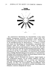
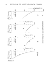
INSTRUMENTAL EVALUATION OF ODOR 243 STERILE SALIVA The sterile saliva was prepared by centrifuging stimulated whole human saliva at 5000 rpm for 10 min, in a refrigerated centrifuge (4øC). The supernatant was passed through a Millipore filter (0.45 3tm) and the flitrate was used for this study. To test the flitrate for sterility, samples were streaked on blood-agar plates and incubated both anaero- bically and aerobically for 24 hr at 37øC. No microbial growth was observed after the period of incubation indicating that the saliva was sterile. DETERMINATION OF ODOR-PRODUCING ABILITY OF MICROORGANISM Each microorganism in sets of six was incubated in a system consisting of 2 ml of sterile saliva, 2 ml of an exogenous substrate, L-cysteine (Nutritional Biochemical Co., Cleveland, Ohio) (5 mg dissolved in 10 ml of sterile saliva) in a Mininert vial (Precision Sampling Corp., Baton Rouge, La.), anaerobically, at 37øC, for a period of 3 hr or 24 hr. After the period of incubation, the headspace above the incubated system was analyzed both organoleptically and instrumentally by gas chromatography and a flame photometric detection system for the presence of the volatile sulfur compounds (VSC) associated with mouth odor. The pH of the incubated system was determined before and after incubation. Sterile saliva and L-cysteine without the organism in phosphate buffer served as controls. Another set of experiments were done adding human blood to the incubation medium to stimulate the growth of the organisms, especially the gram-negatives. GAS-CHROMATOGRAPHY The MT-220 Tracor (Tracor, Inc., Austin, Texas) gas chromatograph equipped with a flame photometric detection system for the sulfur volatiles associated with human mouth odor has previously been described (2, 3). The analyses of the headspace above the incubated systems were obtained at a sensitivity of 1.28 x 10 -•ø ampere full scale (AFS). The sulfur components were separated in a 24 foot x 54 inch (outside diameter) fluorinated ethylene propylene (FEP) column packed with 5% polyphenyl ether and 0.05% phosphoric acid on Chromsorb T (Supelco Inc., Bellefonte, Penn.) (30/60 mesh size). The carrier gas used was ultrapure air (Matheson Gas Products, East Rutherford, New Jersey). The Mininert vial was attached to the inlet of the gas sampling valve leading to the 10-ml sample loop by means of a needle which was put through the septum of the Mininert valve placed in the open position. A 10-ml syringe was attached to the gas sampling valve outlet and the headspace above the incubated system was then allowed to be swept into the sample loop and into the G.C column and the VSC analyzed as previously described (2, 3). RESULTS Figure 1 illustrates the typical chromatogram of the headspace of the incubation system containing a gram-negative microorganism, in this example, Bacteroides melaninogenicus. From comparisons of the retention time with known standards, the
Purchased for the exclusive use of nofirst nolast (unknown)
From: SCC Media Library & Resource Center (library.scconline.org)