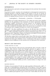
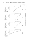
INSTRUMENTAL EVALUATION OF ODOR 243 STERILE SALIVA The sterile saliva was prepared by centrifuging stimulated whole human saliva at 5000 rpm for 10 min, in a refrigerated centrifuge (4øC). The supernatant was passed through a Millipore filter (0.45 3tm) and the flitrate was used for this study. To test the flitrate for sterility, samples were streaked on blood-agar plates and incubated both anaero- bically and aerobically for 24 hr at 37øC. No microbial growth was observed after the period of incubation indicating that the saliva was sterile. DETERMINATION OF ODOR-PRODUCING ABILITY OF MICROORGANISM Each microorganism in sets of six was incubated in a system consisting of 2 ml of sterile saliva, 2 ml of an exogenous substrate, L-cysteine (Nutritional Biochemical Co., Cleveland, Ohio) (5 mg dissolved in 10 ml of sterile saliva) in a Mininert vial (Precision Sampling Corp., Baton Rouge, La.), anaerobically, at 37øC, for a period of 3 hr or 24 hr. After the period of incubation, the headspace above the incubated system was analyzed both organoleptically and instrumentally by gas chromatography and a flame photometric detection system for the presence of the volatile sulfur compounds (VSC) associated with mouth odor. The pH of the incubated system was determined before and after incubation. Sterile saliva and L-cysteine without the organism in phosphate buffer served as controls. Another set of experiments were done adding human blood to the incubation medium to stimulate the growth of the organisms, especially the gram-negatives. GAS-CHROMATOGRAPHY The MT-220 Tracor (Tracor, Inc., Austin, Texas) gas chromatograph equipped with a flame photometric detection system for the sulfur volatiles associated with human mouth odor has previously been described (2, 3). The analyses of the headspace above the incubated systems were obtained at a sensitivity of 1.28 x 10 -•ø ampere full scale (AFS). The sulfur components were separated in a 24 foot x 54 inch (outside diameter) fluorinated ethylene propylene (FEP) column packed with 5% polyphenyl ether and 0.05% phosphoric acid on Chromsorb T (Supelco Inc., Bellefonte, Penn.) (30/60 mesh size). The carrier gas used was ultrapure air (Matheson Gas Products, East Rutherford, New Jersey). The Mininert vial was attached to the inlet of the gas sampling valve leading to the 10-ml sample loop by means of a needle which was put through the septum of the Mininert valve placed in the open position. A 10-ml syringe was attached to the gas sampling valve outlet and the headspace above the incubated system was then allowed to be swept into the sample loop and into the G.C column and the VSC analyzed as previously described (2, 3). RESULTS Figure 1 illustrates the typical chromatogram of the headspace of the incubation system containing a gram-negative microorganism, in this example, Bacteroides melaninogenicus. From comparisons of the retention time with known standards, the

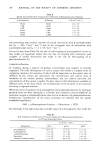
244 JOURNAL OF THE SOCIETY OF COSMETIC CHEMISTS l, H2S 2. CH3 SH :5. {CH3}2 S COLUMN: FEP TEFLON COLUMN PACKED WITH 5 % POLYPHENYL ETHER AND 0.05 % H3PO 4 ON 30/60 MESH CHROMSORB T ATTENUATION = 1.28 x I0 -•ø AFS, TEACOR MT 220 TEMPERATURE = 60 øC CARRIER GAS: AIR Figure 1. GC-FPD chromatogram of headspace of incubated system containing gram-negative microor- ganism. peaks were shown to be that of hydrogen sulfide and methyl mercaptan as major components and dimethyl sulfide as a minor component. The gram-staining character of the microorganisms and the ability to produce VSC from the sterile saliva system with L-cysteine as substrate, in the presence of human blood is shown in Table I. From the table it is clear that only the gram-negatives were capable of producing the volatile sulfur compounds from the sterile incubated system. Veillonella alcalescens, Fusobacterium nucleatum, Bacteroides melaninogenicus and Kleb- siella pneumoniae produced VSC. However, the gram-positives failed to produce VSC. An unpleasant odor was detected organoleptically when VSC was present in the incubated system. Although it is possible to quantitate the amounts of VSC produced, at this time the evaluations were qualitative to determine if indeed the VSC produced Table I Odor and VSC Production by Organisms and Gram Staining Characteristics VSC Odor Gram Organism Production Produced Stain Streptococcus sanguis No None Streptococcus salivarius No None Streptococcus mutans No None Actinomyces naeslundii No None Lactobacillus acidophilus No None Staphylococcus aureus No None Candida albicans No None Bacteroides melaninogenicus Yes Unpleasant Fusobacterium nucleatum Yes Unpleasant Veillonella alcalescens Yes Unpleasant Klebsiella pneumoniae Yes Unpleasant Positive Positive Positive Positive Positive Positive Positive Negative Neganve Negauve Negative Incubation period: 3 hr or 24 hr, 37øC
Purchased for the exclusive use of nofirst nolast (unknown)
From: SCC Media Library & Resource Center (library.scconline.org)