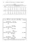
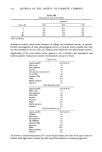
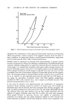
314 JOURNAL OF THE SOCIETY OF COSMETIC CHEMISTS to be less important. Compared to the liver, metabolizing enzymes in the skin are in low concentration and many standard assays for the studies of enzymes in liver are insufficiently sensitive for the study of enzymes in skin (8). Since the finding of novel phototoxic chemicals in fragrance materials is an uncommon event, it is further unlikely that human derreal metabolism will fortuitously act to cause an unexpected phototoxic response. Metabolic participation of the liver in producing phototoxic dermatitis is possible, but the liver would receive dilute quantities of fragrance material substrates. The products of these substrates would then be further subject to excretion by the usual routes, leaving the remainder to be in equilibrium with preferential storage sites. Most of these would be inaccessible to ultraviolet radiation. An additional explanation for the relatively greater sensitivity of the screen compared to intact skin may be attributed to the natural barriers that skin possesses with regard to penetration of test substance or UV light. Alternatively, compromised human skin in which barriers to penetration have been lowered may reduce or even eliminate the differential in sensitivity between the in vitro and in vivo systems. Barriers to penetration in normal skin may be lowered by such factors as disease, advanced age, use of penetrating creams (9), or use of occlusive clothing. It seems appropriate, therefore, that before commercial release of products which have significant photo- toxic activity in vitro, results of tests in humans should be repeated using a suitable model of skin compromised with regard to its resistance to penetration of light and test agent. Lastly, the skin may be less sensitive than the in vitro system in view of the complex series of events which provide the endpoint in vivo. This endpoint, erythema, is a function of dilation of the derreal vascular bed and is in turn dependent on the effects of vasoactive mediators. Such mediators are released following damage (in this case phototoxic damage), and the ability to perceive this effect is subjective compared with the quantitative measurement of zones of inhibition of yeast growth. When recording results in the yeast assay system, it is important to recall that the apparent potency of test materials is a function of diffusion through the agar medium. Evidence of comparable rates of diffusion should be obtained before relative potencies of two materials may be examined. When diffusion becomes a significant concern, fungistatic activity as an indicator of phototoxic events can be monitored in a shaker culture rather than in a semi-solid agar medium. A simple method of measuring light transmittance can provide the measure of light-dependent inhibition. Since diffusion in agar is allowed to progress for 18 hours under UVA exposure, slowly diffusing chemicals should have more than adequate time to migrate, making the use of liquid media rarely necessary. In both the agar and shaker culture method of screening, the dark controls should always be retained. Eugenol, Isoeugenol, methyl salicylate and some of the Coumarin molecules inhibit yeast in the dark as well as in the light. In these cases, a positive phototoxic effect can be declared only when there is a substantial increase in the light mediated inhibition over that in the dark. REFERENCES (1) F. Daniels, Jr., A simple microbiological method for demonstrating phototoxic compounds,J. Invest. Dermatol., 44, 259 (1965).

PHOTOTOXIC ACTIVITY OF FRAGRANCES 315 (2) L. C. Harber and R. L. Baer, Pathogenic mechanisms of drug-induced photosensitivity, J. Invest. Dermatol., 58 (6), 327-342, (1972). (3) M. Bjellerup and H. Moller, Photoaugmentation in drug phototoxicity, J. Invest. Dermatol., 75, 228-229 (1980). (4) S. T. Zaynoun, The quantitative analysis of bergapten in perfumes. J. Soc. Cosmet. Chem., 29, 247-263 (May, 1978). (5) K. H. Kaidbey and A.M. Kligman, "Identification of Contact Photosensitizers by Human Assay," in Current Concepts in Cutaneous Toxicity, Edited by Victor A. Dull and Paul Lazar. (Academic, New York, 1980), pp. 55-68. (6) W. B. Glew, W. P. Roberts, G.I. Malinin and T. P. Nigra, Quantitative determination by bioassay of photoactive 8-methoxypsoralen in serum,J. Invest. Dermatol., 75, 230-234, (1980). (7) K. H. Kaidbey and A.M. Kligman, Photocontact allergy to 6-methylcoumarin, Contact Dermatitis, 4, 277-282, (1978). (8) David R. Bickers, "The Skin as a Site of Drug and Chemical Metabolism," in Current Concepts in Cutaneous Toxicity, Edited by Victor A. Dull and Paul Lazar. (Academic, New York, 1980), pp. 95-126. (9) K. H. Kaidbey and A.M. Kligman, Topical photosensitizers (Influence of vehicles on penetration), Arch. Dermatol., 110, 868-870, (1974).
Purchased for the exclusive use of nofirst nolast (unknown)
From: SCC Media Library & Resource Center (library.scconline.org)