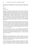
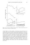
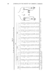
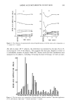
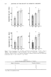
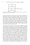
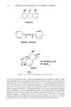
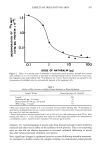
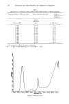
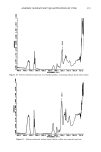
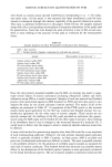
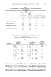
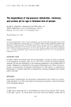
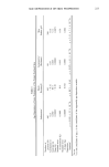
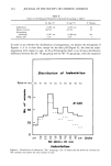
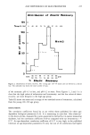
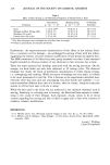
AMINO ACID METABOLITES IN DRY SKIN 193 Table V Levels of Nucleated Horny Cells for Each Skin Type in 77 Subjects Degree of Nucleation* Skin Type A B C 1 2 3 4 5 3 4 2 3 0 0 2 2 10 11 1 0 1 1 4 17 6 1 0 1 1 8 3 2 * See Table I classification scale. metabolism related to keratinization varied in the hyperkeratotic epidermis as shown in Figure 5. Regarding the four compounds mentioned above, His, Glu, Asp, and Orn increased, while their respective metabolites such as UCA, PCA, Ala, and Cit de- creased, suggesting that all the conversions were suppressed when keratinization was pathologically accelerated. The metabolic behavior of each amino acid during a hyper- keratotic period was evaluated in terms of the rate of conversion (Cit/Cit q- Orn, PCA/ PCA q- Glu, Ala/Ala q- Asp, and UCA/UCA q- His). The results are shown in Figure 6. Regarding Cit, which is formed from Arg via Orn, the rate of conversion from Orn to Cit was employed because of the higher value of the correlation coefficient. The rate of conversion for each metabolic pathway was considerably lowered in association with Table VI Amino Acid Metabolism In Vivo METABOLITE METABOLIC PATHWAY PCA • i• :•'l'fi':•'• '- ¾-glutamyI-AA ¾-glutamyl synthetase cyclotrans•erase UCA i'fii'õ'i /NH, : ........... =- :UCA! histidase carbamoyl Orn , ....... ! •,Urea phosphat_,..•½_ Pi Cit --i'b•:•i -- • ' ......
!Arg. ' : ........ ornithine ........ arginase carbamoyltransferase COz Ala / aspartate 4-decarboxylase , ,

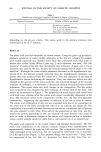
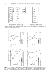
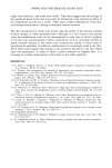
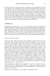
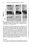
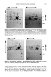
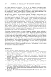
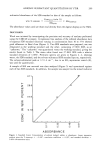
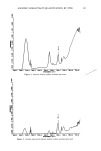
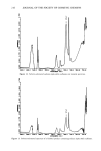
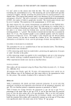
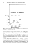
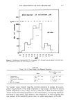
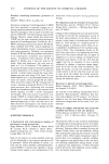
194 JOURNAL OF THE SOCIETY OF COSMETIC CHEMISTS -0.75 GLU PC^ -0.46 HIS uc^ -0.31 -0.54 ARG = ORN CIT -0.75 ASP ALA Figure 8. Correlation coefficients showing the inverse relationships between levels of free amino acids and their metabolites in cheek skin of 46 female subjects. acceleration of keratinization. A similar tendency was observed when hexadecane and ultraviolet rays as well as LAS were applied. In addition, the rates fell further as the irradiation with ultraviolet rays was prolonged (9). These suggest that the conversion rates well reflect the degree of hyperkeratosis. The decreases in the rate of conversion may be attributed to a decrease in activity of enzymes which catalyze the conversion or a scarcity of time for relevant metabolism due to acceleration of the turnover. The precise mechanism, however, remains as yet to be elucidated. In hyperkeratotic epi- dermis, at least, free amino acids in the stratum corneum are affected, and the rates of amino acid conversion through the four metabolic pathways involved in the keratini- zation especially reflect this feature of keratinization. Taking advantage of the process, the skin condition could biochemically be estimated with water soluble constituents extracted from the skin surface without invasion. Dry skin in winter. Each of the four amino acid conversion rates measured in dry skin was lower than those in normal skin (p 0.05), as shown in Figure 7. This suggests that keratinization was disordered simultaneously with the skin's getting chapped. Para- keratotic changes in the epidermis can be examined by usual histological techniques. It is, however, difficult to perform biopsies on many subjects. A new technique reported recently provides a simple method of estimating parakeratotic changes, wherein residual nuclei in the stratum corneum are counted in a cornified cell specimen detached with the aid of adhesive tape (10, 11). In the investigation of parakeratotic changes, the residual nuclei were evaluated by the new procedure. The results are shown in Table V. In normal keratinization, the nuclei disappear in the process of differentiation, and the stratum corneum consists of non-nucleated cells alone. When the keratinization is disturbed, the disappearance becomes incomplete, resulting in nucleated cells in the stratum corneum. Although all the subjects chosen for the present study were healthy, only 19.5% showed stratum corneum consisting of non-nucleated cells alone. Accord- ingly the nucleated horny cells were found in a considerable portion of the subjects. The degree of parakeratosis was proportional to the roughness of the skin. As described above, the studies on changes in the composition of free amino acids in the stratum corneum and the extent of parakeratosis suggest changes in the epidermis in association with skin roughness and that skin lesions associated with dry skin do not stay within the stratum corneum. In experimentally induced hyperkeratosis, the skin surface ex- hibited changes similar to the surface of dry skin: the surface became dry, furrows and
Purchased for the exclusive use of nofirst nolast (unknown)
From: SCC Media Library & Resource Center (library.scconline.org)