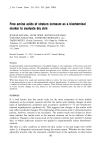
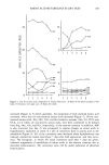
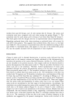
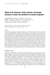
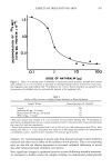
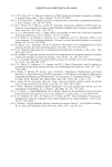
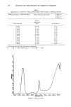
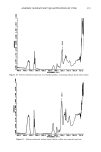
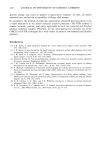
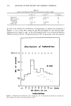
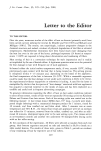
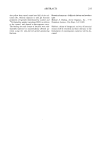
198 JOURNAL OF THE SOCIETY OF COSMETIC CHEMISTS Anthralin Benzoyl Peroxide OR • '-,•OR = • R 1= CO-(CH= )•=-CH 3 O R =.- COCH= CH=OH TPA Figure 1. Structures of TPA, anthralin, and benzoyl peroxide. of TPA to mouse skin causes a series of morphologic and biochemical changes. These include inflammation and epidermal hyperplasia as well as changes in cell growth and differentiation (7,8). Anthralin has been shown to be the most potent non-phorbol ester tumor promoter in the mouse skin bioassay (4). It inhibits DNA replication and repair (9), mitotic activity in skin (10), and DNA, RNA, and protein synthesis in cultured human fibroblasts (11). Benzoyl peroxide is a free radical-generating compound that, in addition to its tumor-promoting activity, is a potent inducer of epidermal hyperplasia (12). Previous studies have shown that benzoyl peroxide can induce mor- phologic changes in mouse skin similar to those observed with TPA (3). Benzoyl peroxide has also been shown to decrease cellular metabolic cooperation in Chinese hamster cells in vitro, a property correlated with tumor-promoting activity (3).

EFFECTS OF IRRITANTS ON SKIN 199 In previous studies we analyzed the effects of treatment of mouse epidermis with TPA on the profile of newly synthesized proteins using two-dimensional polyacrylamide gel electrophoresis (13). Over 200 individual proteins were resolved in acidic gels and at least 10 of these showed major (five-fold or more) increases or decreases in response to TPA. Several of these proteins appeared to be keratins which are important markers of epidermal cell differentiation. The present study was designed to investigate whether the tumor promoters anthralin and benzoyl peroxide produced similar changes in the production of mouse epidermal proteins. Our results demonstrate that these compounds produce distinct effects on epidermal protein synthesis, suggesting that there may be several alternate mechanisms of tumor promotion in the mouse skin. EXPERIMENTAL Animals used were female, CD-t mice 7-t0 weeks of age, supplied by Charles River Breeding Laboratories. Anthralin was provided by Elder Pharmaceuticals. Benzoyl per- oxide was purchased from Aldrich. TPA was obtained from Consolidated Midland Corp. Each chemical was dissolved in acetone (Fischer) and freshly prepared for each experi- ment. 35S-methionine (tOO0 Ci/mmole) was purchased from New England Nuclear. Ampholines for isoelectric focusing gels were from LKB, Sweden. Tissue culture me- dium and newborn calf serum were obtained from Grand Island Biological Company. LABELING OF EXCISED EPIDERMIS Twenty-four hours before application of the compounds the dorsal skins of the mice were shaved. TPA, anthralin, and benzoyl peroxide were applied in 0.2 ml of acetone directly to the shaved skin. Control mice received 0.2 ml of acetone alone. Twenty- four hours after treatment, the mice were sacrificed by cervical dislocation, and a chemical depilatory (Neet, Whitehall Laboratories) was applied for 3 minutes. The depilatory was removed from the skin by washing the animal under cold water. The skin was then excised and the subcutaneous adipose tissue was removed with a scalpel. The sample was cut into 4 sections (approx. 2 cm 2 each) and floated dermal side down in 3 ml of incubation medium in 5 cm plastic tissue culture dishes. The incubation medium consisted of Dulbecco's Modified Eagle's Medium, without methionine, sup- plemented with 5% newborn calf serum and tOO •Ci/ml 35S-methionine. The culture dishes were then incubated at 37øC in a humidified CO2 incubator. After 4 hours, pure epidermis was harvested by the heat treatment method of Raineri et al. (14) and immediately placed in 0.4 ml modified O'Farrell lysis buffer (15) consisting of 9.5 M urea, 2% w/v Nonidet P 40, and 5% 2-mercaptoethanol. The resulting lysate was sonicated for t minute on a Sonic Dismembrator (Artek Corp., Model 150). Samples were then stored at -70øC until use. For protein analysis, samples were centrifuged for 5 minutes in a Fischer micro-cen- trifuge (model 235) at approximately 10,000 x g. Only clear supernatant fractions .were used. Protein was determined by the method of Bradford (16), using bovine serum albumin as the standard. Radioactivity was measured on a Tracor Mark III scintillation counter.
Purchased for the exclusive use of nofirst nolast (unknown)
From: SCC Media Library & Resource Center (library.scconline.org)