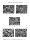
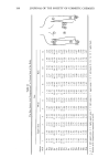
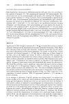
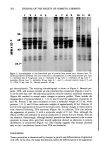
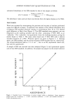
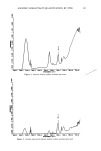
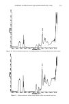
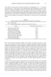
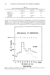
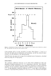
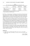
200 JOURNAL OF THE SOCIETY OF COSMETIC CHEMISTS POLYACRYLAMIDE GEL ELECTROPHORESIS One-dimensional discontinuous SDS-polyacrylamide slab gels were run according to the method of Laemmli (17). The separating gel contained 10% polyacrylamide. Du- plicate gels were run for autoradiography and protein silver staining (18). Samples for silver staining contained 10-50 p,g of protein, and for autoradiography, approximately 100,000 cpm. Two-dimensional polyacrylamide gel electrophoresis with isoelectric focusing was carried out as described by O'Farrell (15) with some modifications. Am- pholines, pH 5-8 and pH 3.5-10, were used in the equilibrium isoelectric focusing dimension, and discontinuous 10% polyacrylamide gels were used in the second di- mension. 1.6% ampholines (pH 5-8) and 0.4% ampholines (pH 3.5-10) were added to the epidermal samples prior to first dimension electrophoresis. Pre-stained molecular weight standards (Bethesda Research Labs.) used were cytochrome C (molecular weight 12.3 kd), [3-1actoglobulin (18.4 kd), tx-chymotrypsinogen (25.7 kd), ovalbumin (43 kd), bovine serum albumin (68 kd), phosphorylase B (92.5 kd), and myosin H-chain (200 kd). Gels were fluorographed using EnHance (New England Nuclear) to visualize radioactive peptides. RESULTS Application of TPA (10 •g) or anthralin (0.1-80 •g) to mouse skin resulted in marked irritation characterized by hyperemia and increased epidermal thickening. These effects were observed within 24 hours after treatment. Anthralin-treated skin was also discol- ored with a characteristic yellow-brown appearance. In contrast, we found that 24-hour treatment with benzoyl peroxide (1-40 mg) did not induce any of these changes in the skin, even at the highest dose tested. In order to determine if the increased epi- dermal thickening was associated with altered protein production, we examined the effects of these compounds on epidermal protein synthesis. Skin fragments were pulse- labeled with 35S-methionine and extracts prepared as described in the Experimental section. Aliquots of these extracts were acid precipitated and counted for radioactivity. We found that anthralin caused a significant, dose-related decrease in protein synthesis in the range of 0.1 to 80 •g/mouse (Figure 2, Table I). At 80 }xg, the maximum dose tested, protein synthesis was inhibited by 78%. This dose of anthralin corresponds to the optimal concentration required for tumor-promotion in mouse skin (4). In contrast, with tumor-promoting doses of benzoyl peroxide (40 mg, see Table I) there was a 15% stimulation of protein synthesis. TPA (10 •g/mouse) produced a 37% decrease in protein synthesis. In an attempt to determine if specific proteins were being altered, we analyzed the protein profiles of the control and treated epidermis using one-dimensional polyacryl- amide gel electrophoresis. Parallel gels were run and stained with silver for analysis of total protein content or subjected to autoradiography for determination of newly syn- thesized proteins (see Experimental section). Silver-stained gels revealed that treatment of mouse skin with promoting agents produced no significant effects on protein dis- tribution (not shown). Autoradiographs of these gels, however, revealed several major differences between anthralin and benzoyl peroxide (Figure 3). Anthralin (40-80 •g) treatment induced the disappearance of a protein with a molecular weight of approxi- mately 60 kd, and the appearance of several proteins in the molecular weight range of 45-50 kd. These findings are similar to those observed with TPA (Figure 3 and

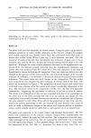
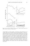
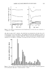
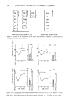
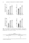
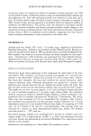
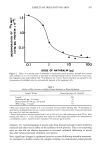
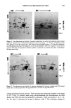
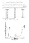
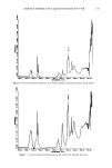
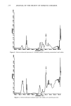
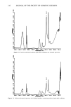
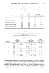
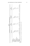
EFFECTS OF IRRITANTS ON SKIN 201 0.1 I I 100 DOSE OF ANTHRALIN (•g) Figure 2. Effect of increasing doses of anthralin on epidermal protein synthesis. Animals were treated with anthralin in 0.2 ml of acetone as described in the Experimental section. Twenty-four hours later, skin fragments were pulse-labeled with 35S-methionine for 4 hours. Protein synthesis was assessed by the incorporation of radiolabel into the acid-insoluble material of the epidermal cells. Table I Ability of Skin Irritants to Inhibit Protein Synthesis in Mouse Epidermis Sample a (Dose) Protein Synthesis (% Control) b Control Anthralin (80 Ixg, 354 nmol) Benzoyl peroxide (40 mg, 165 Ixmol) TPA (10 Ixg, 17 nmol) 100 22 115 63 a Mice were treated with irritants in 0.2 ml of acetone 24 hours before labeling with 35S-methionine as described in the Experimental section. Control animals received 0.2 ml acetone alone. Each point represents the mean of at least four experiments. b Protein from extracts was precipitated with 3 ml of 10% (w/v) ice cold trichloroacetic acid (TCA). After rinsing with TCA (2 X 3 ml), precipitates were collected on filter paper and radioactivity determined on a scintillation counter. Control extracts contained 740 cpm per }.tg of protein. reference I3). Autoradiographs of protein gels from benzoyl peroxide-treated epidermis indicated that there was no effect on the synthesis of new proteins. This is not surprising since we also did not observe hyperemia or increased epidermal thickening in mouse skin after benzoyl peroxide treatment (not shown). Since significant changes in epidermal proteins occurred following anthralin treatment, we attempted to further resolve the samples utilizing two-dimensional polyacrylamide
Purchased for the exclusive use of nofirst nolast (unknown)
From: SCC Media Library & Resource Center (library.scconline.org)