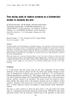
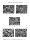
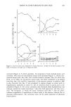
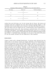
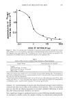
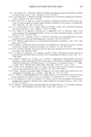
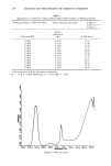
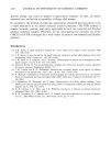
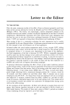
AMINO ACID METABOLITES IN DRY SKIN 185 ..' ! 4. 5 Figure 1. Typical skin surface conditions classified by replica method. Photos 1 and 2 represent dry skin. Photos 4 and 5 represent normal skin. Photo 3 represents an intermediate skin condition. (x 25)..

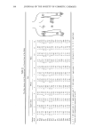
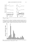
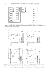
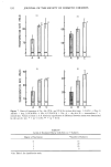
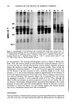
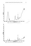
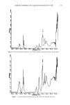
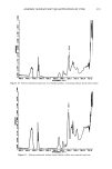
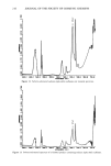
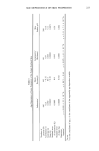
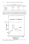
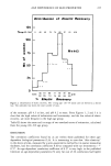
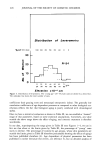
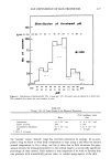
186 JOURNAL OF THE SOCIETY OF COSMETIC CHEMISTS Table I Classification of Stripped Cornified Cells Based on Degree of Nucleation Degree of Nucleation Number of Nuclei per Sample No nucleus is present One or two nuclei are observed 3-5 nuclei are observed More than 6 nuclei are observed depending on the pre-set criteria. Free amino acids in the stratum corneum were determined in 46 of 77 subjects. RESULTS Free amino adds and their metabolites in stratum corneum. Using the glass cup procedure, adequate quantities of water soluble substances in the stratum corneum for analysis were readily extracted into distilled water from skin pretreated with ethyl ether to remove skin surface lipids. When a glass cup, 2 cm in diameter, was used, 100-200 nmol/cm 2 of amino acids and their metabolites were extracted. A glass cup, 4 cm in diameter, was used for the face, because the extracted amount was as small as 30-100 nmol/cm 2. Although the water soluble substances decreased in the hyperkeratotic epi- dermis (4,6), the amount actually extracted from the hyperkeratotic epidermis was greater than that extracted from the control skin. This was explained on the basis of hyperkeratotic lesions facilitating the extraction. Thus, the extr-action reflects not only changes in the amount of free amino acids but also structural changes of the stratum corneum. Accordingly, it is irrelevant to discuss the amou•nt of extracted water soluble substances. This report deals only with changes in the composition. The free amino acid composition was measured by this technique at various sites of the body. The results are summarized in Figure 2. The proportions of minor amino acids varied little with body sites, whereas other free amino acids varied considerably depending on the sites. The variations noted in the composition of Orn, Cit, and Arg were especially characteristic, suggesting the importance of selection of the site to serve as control. The amino acid composition at one site was compared with that at its counterpart on the other side of the body (contralateral site) or at an adjacent site (Table II). The composition is practically the same between one site and its contralateral site or between two sites adjacent to each other. Even each of the proportions of Arg, Orn, and Cit, which vary greatly depending on site, are virtually the same between such a pair of sites. In order to determine changes in free amino acid composition due to hyperker- atosis, control samples should be taken from a contralateral site or an adjacent site. Free amino acid composition in the stratum corneum and abnormal keratinization. The free amino acid composition in the stratum corneum changed when hyperkeratosis was experimentally induced by applying LAS solution. The composition remained essen- tially unchanged at the control site, but the composition varied from 3 to 21 days after the LAS application (Figure 3). The greatest change was noticed after 7 days, when the proportions of Ser, Cit, Ala, etc. decreased, but those of Asp, Glu, Orn, etc.
Purchased for the exclusive use of nofirst nolast (unknown)
From: SCC Media Library & Resource Center (library.scconline.org)