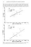
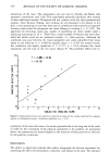
j. Soc. Cosmet. Chem., 35, 327-338 (September/October 1984) Evaluation of hydration state and surface defects in the stratum corneum: Comparison of computer analysis and visual appraisal of positive replicas of human skin RICHARD I. MURAHATA, DANIEL M. CROWE, and JOHN R. ROHEIM, Armour-Dial, Inc., Armour Research Center, 15101 N. Scottsdale Road, Scottsdale, AZ 85260. Received March 23, 1984. Synopsis This study was designed to develop a noninvasive model system for analyzing the condition of human skin in vivo and to provide a method of objective measurement which would correlate with the subjective evaluation of the skin surface. Positive replicas of the upper arm were prepared for scanning electron microscopy and surfanalysis. Skin condition was evaluated by scoring SEM photomicrographs for plump- ness, an indication of hydradon, and for stratum comeurn damage represented by surface scales and cracks. Skin surface profiles parallel with and perpendicular to the major furrows were produced using a surfan- alyzer. The profiles were computer analyzed for standard roughness parameters utilized by the metals industry, including arithmetic average roughness (Ra) and mean depth of roughness (Rz). Eleven to thirteen individual samples were examined. The subjective value for plumpness correlated (p 0.02) with both Ra and Rz, and the correlation was not dependent on the trace direction of the surface profile. The subjective evaluation of stratum corneum damage, however, did not correlate with any of the roughness parameters or with plumpness. This may be due in part to the sensitivity of the measurement, with cracking and flaking being restricted to small changes in the upper few microns of stratum corneum and plumpness being a reflection of larger changes throughout the stratum corneum and perhaps involving the epidermis. A special program was written which could identify cracks in a roughness profile with a high degree of certainty, and the number of cracks identified was significantly correlated (p 0.01) with a subjective assessment of the corresponding photomicrographs. This technique provides a powerful tool for assessing skin condition and the effects of cosmetic agents on the topography of the stratum corneum. INTRODUCTION The ability to evaluate skin surface characteristics in an objective, noninvasive manner can result in a better understanding of perceived properties of the skin (e.g., smooth- ness) and in the development of more efficacious cosmetic products. Several methods have been used to evaluate surface texture including stereomicroscopy (1), macropho- tography (2,3), ellipsometry (4), and skin friction (5,6). Surfanalysis or profilometry (7-12) is probably the most well established, historically having been used by the metals industry three decades before being adopted by researchers studying skin surface morphology. Surfanalysis utilizes a stylus instrument to generate profiles of positive 327

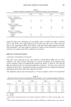
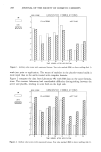
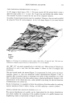
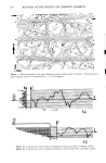
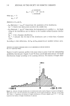
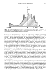
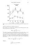
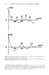
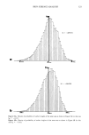
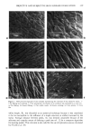
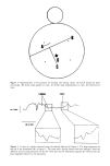
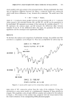
328 JOURNAL OF THE SOCIETY OF COSMETIC CHEMISTS skin replicas, and several profile parameters to quantitate deviations from a flat surface have been standardized by the American Society of Mechanical Engineers (13). In this investigation, skin surface replicas produced by an accurate and precise technique (14) were examined by scanning electron microscopy (SEM) and surfanalysis. The pho- tomicrographs were evaluated subjectively for "plumpness" as an indication of hydration and moisturization (15-17) and for surface defects such as cracks and desquamating sheets (flakes). A significant correlation was observed when SEM evaluations for plump- ness were compared with computer-generated roughness parameters derived from sur- fanalysis profiles. Cracks were identified in surfanalysis profiles and described mathe- matically through an iterative process comparing computer analysis of traces with visual subjective evaluation of photomicrographs. Computer-assisted surfanalysis provides a noninvasive objective evaluation that correlates with a subjective visual assessment of the skin surface condition. An added benefit to this method is the creation of permanent records which can be referenced at a later date. MATERIALS AND METHODS SUBJECTS Thirteen volunteers without apparent skin disease were used in this study. Replicas were made of a small area on the extensor aspect of the upper arm. REPLICA PREPARATION AND EVALUATION A negative replica of the site was generated using a silicon based dental impression material. A polyethylene cast (positive replica) was prepared from the negative skin impression. The development of this methodology was the subject of a previous com- munication (14). Although the fidelity of using positive casts has been questioned (10), preliminary work with the technique (14) indicated that repeated replicas from the same site were not significantly different at the current level of sensitivity. The cast was pegged, gold coated, and photographed at 25 ) and 125 ) magnification by scanning electron microscopy (AMR 1000A). The photomicrographs were scored sub- jectively for "plumpness" (moisturization), "flaking" (uplifted plates and desquamating sheets), and cracking (Figure 1). The surface profile of the cast was traced with a surfanalyzer (Federal Products System 2000) which computes, displays, and records the profile using a linear drive unit equipped with a 0.0001-inch radius diamond stylus. The cast was placed on a translational/rotational stage and a 7.5 mm-trace made parallel with and perpendicular to the principal furrows of the skin. Four replicas were traced six times in each direction, and greater than 75% of the values for Ra and Rz fell within the 95% confidence limits of the mean. A single trace in each direction was used thereafter because the error introduced by hitting a bead of perspiration, hair follicle, or other artifact was greater than the intrasample variability. The reproduc- ibility of a given scan was greater than 99%. The data were collected as a two- dimensional array of 1350 points and were analyzed with the aid of a MINC 11 minicomputer (Digital Equipment Corporation). Several methods of profile analysis devised by the metals industry were examined for their utility in examining skin surface replicas. Some of the methods explored are illustrated in Figure 2. Maximum peak-to-
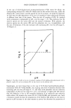
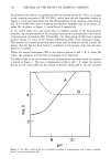
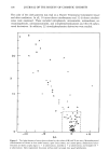
Purchased for the exclusive use of nofirst nolast (unknown)
From: SCC Media Library & Resource Center (library.scconline.org)