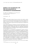
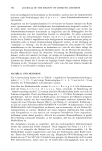
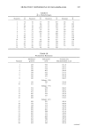
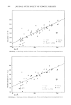
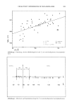
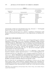
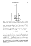
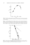
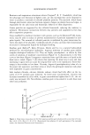
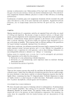
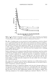
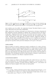
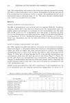
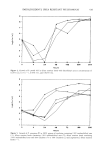
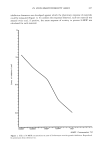
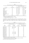
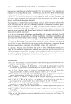
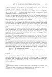
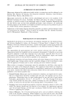
426 JOURNAL OF THE SOCIETY OF COSMETIC CHEMISTS This paper presents data on a modification of the exposure time for the quantitative i, vitro assay reported by Tenenbaum eta/. (2) and the assessment of low level activity for a number of fragrance oil materials previously reported. In addition, an investigation of "quenching," the potential to inhibit a phototoxic response, was conducted by incor- porating benzophenone and sunscreen agents into the standard methanol vehicle. The results from these procedures were compared to published animal and human data. METHODS LIGHT SOURCE/ULTRAVIOLET EMISSION READINGS A bank of four black light tubes (Westinghouse F40BL, 320-400 nm, peak 350 nm) was utilized for all assays. Ultraviolet intensity was measured using a long-wave J221 Black-Ray meter (Ultraviolet Products, Inc.). Energy readings for animal assays ranged from 1.2 to 1.8 mW/cm 2. Flux tbr the in vitro assay ranged from 1.0 to 1.2 mW/cm 2 for the 18-hour assay and 2.0 to 2.5 mW/cm 2 for the 7-hour assay. UV emissions for the in vitro assays were taken through the top of a petri dish (Falcon, No. 1029) which simulated the amount of UV energy reaching the test sites. TEST MATERIALS A 5% test material concentration was used for in vitro studies based on preliminary work which demonstrated that higher concentrations of the fragrance materials elicited antimicrobial activity, limiting the ability of the assay to evaluate the phototoxic po- tential of these materials. A 10-fold exaggeration of the in vitro concentration was used for in'vivo testing based on the greater degree of sensitivity exhibited by the yeast assay compared to the animal model. 18-HOUR IN VITRO ASSAY Saccharomyces cerevisiae (isolated from Fleishman's Yeast) was used in an agar overlay technique wherein 3 ml of a 1% suspension of yeast (approximately 1.7 X 10⁵ or- ganisms/ml) in molten plate count agar (Difco Laboratories No. 0479-01) was added to a 20-ml agar plate. Quarter-inch blank paper discs (Baltimore Biological Laboratory, No. 31039) were impregnated with 25 ptl of the test compound dissolved in methanol and were allowed to dry for 15 minutes at room temperature prior to being placed on the surface of the agar plate. Two plates containing test materials were exposed to UVA (Westinghouse F40BL tubes) for 18 hours at a distance of 31 cm and then incubated at 31-35øC for 48 hours. An additional plate, not irradiated but incubated for 48 hours, served as a control. Phototoxic materials were identified by a clear zone of growth inhibition in the irra- diated plates and none in the non-irradiated plate. A negative phototoxic response is indicated by absence of growth inhibition on both plates. The diameter of the zone of inhibition was measured by an antibiotic zone reader (Fisher-Lilly, Inc.). To quantify the phototoxic activity of the test materials, a curve of 8-Methoxy psoralen concentrations (ICN Pharmaceuticals, Inc. Lot #103296), ranging from 0.01% to 0.0001% (with the latter being the lower limit of detection for the assay) versus zone of

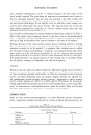
IN VITRO PHOTOTOXICITY ASSAY 427 inhibition diameters was developed against which the phototoxic response of materials could be measured (Figure 1). To confirm the responses observed, each test material was assayed twice and, if positive, the mean response of activity in percent 8-MOP was calculated for each material. ' b.cx5os''' 'o.do•'' 'o.dos''' 'o.&' ' ' ' ' ' ' 8-MOP Concentration (%) Figure 1. Effect of 8-MOP concentration on zone of Saccharomyces cerevisiae growth inhibition. Reproduced by permission from reference (2).
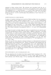
Purchased for the exclusive use of nofirst nolast (unknown)
From: SCC Media Library & Resource Center (library.scconline.org)