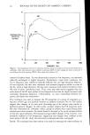
STRATUM CORNEUM HYDRATION 13 shown altered water loss with changing ambient RH (6, 15,19-23). It is interesting to compare results obtained by Blank using two different techniques to measure flux. By measuring water loss through SC in contact with an aqueous buffer on the inner surface, he showed a decrease in flux with increasing ambient RH (6). In these experiments, the inner surface of the SC was maintained at a constant water content while the outer surface was in equilibrium with ambient conditions. Thus, as the ambient RH in- creased, the water concentration gradient, and hence flux, decreased. More recently, Blank and co-workers (18) measured the flux of radiolabelled water vapor through SC equilibrated at various relative humidities and showed increasing flux with increasing RH. In these experiments, a constant gradient of tritiated water was maintained across the SC and, thus, the increase in flux with RH reflected an increase in the diffusion and partition coefficients with increasing water content of the SC. From measurements of in vitro kinetics of water sorption and desorption in the SC as a function of RH, EI-Shimi and Princen (12) also measured changed in the diffusion coefficient with water content. Like Blank eta/. (18), their results showed increasing diffusion coefficient with increasing water content, approaching a constant value at a water concentration near 0.3 g/cm 3. Together, these results show that changes in am- bient RH can alter flux by two opposing mechanisms. The flux is determined by the net effect of the diffusion and partition coefficients, which increase with RH, and the gradient, which decreases with RH. As discussed by Miller eta/. (24), these opposing mechanisms may result in a water flux maximum at intermediate RH, with lower values at both higher and lower RH. The thickness of the SC also affects the water gradient and, in turn, the flux. For example, Blank (15) measured water flux through callus removed from the foot as a function of sample thickness. His results showed that the flux increased with decreased thickness of the sample. Since the inner and outer surfaces of the sample were main- tained at constant water concentrations, decreasing the thickness resulted in an in- creased gradient and, hence, flux. In addition, the SC increases in thickness when hydrated and, thus, as noted by numerous investigators (11,15,25), changes in flux must be corrected for water-induced swelling of the SC. Unfortunately, these correc- tions often are not possible (especially in vivo), since precise swelling changes cannot be measured. Differences in in vitro flux were also seen for samples from different sites on the body. For example, Blank (15) showed that the in vitro water flux through plantar callus was greater than through abdominal skin, even though the SC in the latter tissue was much thinner. Extrapolation of flux-vs.-thickness data for callus samples to a thickness com- parable to SC in the abdomen yielded a hundred-fold greater flux in plantar tissue. Similarly, Smith eta/. (17) showed that in vitro flux through scrotal skin was about 20-fold greater than through abdominal skin, even though both have SC of comparable thickness. Finally, Scheuplein and Blank (26) showed that, while the flux at eight sites on the body varied by an order of magnitude, the water diffusion coefficients varied by three orders of magnitude. These results demonstrate inherently different diffusional resistance at various skin sites. Several investigators have examined the role of lipids in SC water barrier properties. For example, while treatment of the skin surface with 6M urea, 0.1 M NaOH, and boiling water had little effect on water loss (16), extraction of the SC with lipid solvents dra- matically increased the in vitro flux, and replacement of the extracted lipids restored the

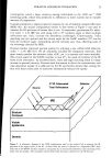
14 JOURNAL OF THE SOCIETY OF COSMETIC CHEMISTS barrier function almost entirely (6,16,27). In addition, variation in in vitro water flux between abdominal and leg SC has been related to differences in SC lipid content be- tween the sites (28). Other results, supporting the important role of lipids in SC water barrier function, are outlined by Elias (29). In summary, in vitro results show that water loss measurements reflect the integrity of the SC barrier. Furthermore, changes in SC water content, thickness, and lipid content, as well as sample site, can dramatically alter water loss values. IN VIVO TECHNIQUES TRANSEPIDERMAL WATER LOSS (TWL) An excellent review of early in vivo measurements of TWL was published by Idson (30). A good review of the general principles of TWL measure was also recently published (24). Techniques to measure TWL in vivo generally fall into two categories, ventilated and unventilated. In the former, a continuous flow of carrier gas passes through a chamber attached to the surface of the skin and changes in water content of the gas are monitored either gravimetrically (31) or by a variety of other sensing techniques, including thermal conductivity (32) and electrohygrometry (33). Ventilated techniques suffer from the fact that the stream of carrier gas over the skin surface can create conditions (e.g. evaporative cooling) different from ambient conditions. In contrast, unventilated techniques use an open chamber placed on the skin surface. Water loss through the skin then alters the RH within the chamber and, thus, measurement of changes in moisture within the chamber provides a measure of TWL. Difficulties with this technique arise when rapid changes in ambient conditions and air movement alter the RH within the open chamber. As with in vitro water flux measurements, TWL measurements in vivo are dependent on a number of factors. For example, the water content of the SC can dramatically alter TWL. Maibach and co-workers measured TWL on the forearms of 10 volunteers and found that the flux increased by about four-fold after several days of continuous occlu- sion but returned to normal 18 hrs after removal of the occlusive film (34). Since water content of the SC increases with occlusion, these results suggest, in agreement with in vitro results, that increased hydration results in increased flux. Similarly, Cooper and van Duzee (35) have noted that while occlusive agents (e.g. petrolatum) applied to the skin cause a transient decrease in TWL, several hours after application (and presumably after at least partial removal of the occlusive agent) the TWL was greater than the pre-treatment values. The increase in water loss was explained by an increase in the diffusion coefficient of water as the SC was hydrated. These authors also point out that good moisturizing agents may actually increase TWL through a hydration-induced in- crease in the diffusion coefficient, rather than the traditionally held belief that such agents seal the barrier and, hence, lower TWL. These results serve to once again under- score the complex relationship between water content and flux in the SC. The in vivo water flux can also be altered by changes in skin temperature. For example, Spruit and Herweyer (36) noted an approximate two-fold increase in TWL at several skin sites with an increase in skin temperature from 23 to 33øC. TWL was also mea- sured on surgery patients under induced hypothermia with results again showing an approximate two-fold increase with an increase in skin temperature from about 27 to 34øC (37). Similarly, an average two-fold increase in TWL was noted at four different
Purchased for the exclusive use of nofirst nolast (unknown)
From: SCC Media Library & Resource Center (library.scconline.org)