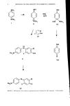
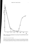
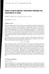
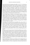
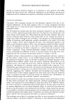
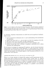
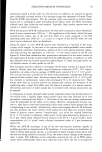
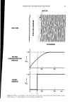
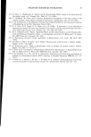
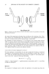
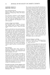
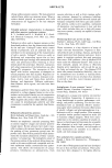
STRATUM CORNEUM HYDRATION 29 SECTION uJ z o WATER :*..:.'*...,•:..y.' :::..'*:lit*.*.':: .*.v. ,Y:' ::**':..* ?, **:**"**• .'.',•, **:.•:..' .::• .*. ., ': EPIDERMIS WATER CONCENTRATION PROFILE MODEL T PROFILE Cw 1.0 1.0 I 4O I I 20 40 •m Figure 8. Water concentration in the outer layers of the skin experimental and model profiles. Taken from reference 77 with permission of the authors and MTP Press.

30 JOURNAL OF THE SOCIETY OF COSMETIC CHEMISTS CONCLUSION The measurement of water in the SC has progressed from in vitro investigations, the results of which are important in understanding the mechanisms of binding and flux, to the more clinically relevant in vivo techniques. In particular, in vitro results have shown that the mechanism of water binding changes over the range of about 0.1 to 0.5 (w/w) water content. At low hydration, water is tightly bound to polar groups of SC constit- uents (i.e., proteins), while at higher hydration, lower energy water-water binding occurs. Similarly, water flux changes with hy. dration over the same range of water content due to changes in D, Km, and dc/dx. Results from in vivo investigations have shown that water content of the SC increases continuously with increasing depth, again spanning the range from about 0.1 to 0.5 (w/w). Thus, the mechanisms of flux and binding discovered with in vitro techniques are revelant to in vivo tissue. Recent em- phasis with in vivo techniques has been directed toward quantitative measurement of the SC water profile. With further sophistication, it should be possible to routinely mea- sure SC hydration in the clinic and thereby relate dermatological disorders (i.e., psori- asis, ichthyosis, etc.) to altered mechanisms of water uptake and flux. ACKNOWLEDGEMENTS The author would like to thank Dr. James E. McKie for his help in reviewing the manuscript, and Drs. McKie and Steven L. Jacques for many enlightening discussions on this subject. (6) (7) (8) (9) (10) (11) REFERENCES (1) R. M. Dahlgren and W. H. Elsnau, Measurement of skin condition by sonic velocity,J. Soc. Cosmet. Chem. 35, 1-20 (1984). (2) P. Flesch and E. C. Jackson Esoda, Deficient water-binding in pathologic horny layers, J. Invest. Dermatol. 28, 5-13 (1957). (3) H. Tagami, Y. Kanamuru, K. Inoue, S. Suehisa, F. Inoue, K. Iwatsuki, K. Yoshikuni, and M. Yamada, Water sorption-desorption test of the skin in vivo for functional assessment of the stratum corneum, J. Invest. Dermatol., 78, 425-428 (1982). (4) P. Frost, G. D. Weinstein, J. W. Bothwell, and R. Wildnauer, Ichthyosiform dermatoses. III. Studies of transepidermal water loss, Arch. Dermatol., 98, 230-233 (1968). (5) M. Gloor, B. Heymann, and T. Stuhlert, Infrared-spectroscopic determination of the water content of the horny layer in healthy subjects and in patients suffering from atopic dermatitis, Arch. Dermatol. Res., 271, 429-436 (1981). I. H. Blank, Factors which influence the water content of the stratum corneum, J. Invest. Dermatol., 18, 433-440 (1952). R. L. Anderson, J. M. Cassidy, J. R. Hansen, and W. Yellin, Hydration of stratum corneum, Bio- polymers, 12, 2789-2802 (1973). T. S. Spencer, C. E. Linamen, W. A. Akers, and H. E. Jones, Temperature dependence of water content of stratum corneum, BritishJ. Dermatol., 93, 159-164 (1975). B. F. Van Duzee, The influence of water content, chemical treatment and temperature on the rheo- logical properties of stratum corneum, J. Invest. Dermatol., 71, 140-144 (1978). B. A. Turek, M. Vieu, M. Leduc, I. M. Nadvornik, and E. Jedla, Water content and moisture- binding capacity of some types of human soft and hard horn, Curt. Med. Res. Opinion., 7, 87-90 (1982). A. F. El-Shimi and H. M. Princen, Water vapor sorption and desorption behavior of some keratins, Colliod and Polymer Sci., 256, 105-114 (1978).
Purchased for the exclusive use of nofirst nolast (unknown)
From: SCC Media Library & Resource Center (library.scconline.org)