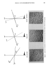
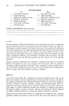
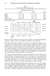
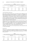
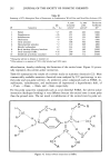
146 JOURNAL OF THE SOCIETY OF COSMETIC CHEMISTS The surface of human skin is traversed by parallel primary and secondary furrows (2) whose intersections create distinctive triangular and rectangular patterns, peculiar to each skin region. These constitute the skin's microrelief, a topographic map of plateaus and valleys. Several early studies on skin surface describe the microrelief of aging skin (3,4). In actinically damaged skin, such as the dorsum of the hand, this microrelief becomes blurred and the fine markings effaced altogether (4). In a previous paper, we used Image Analysis of skin replicas to follow the age-associated evolution of the major furrows of forearm skin (5). We found an age-associated decrease in the density of the primary furrows, accompanied by an increase in their depth. Deeper furrows cause an increase in total surface area, which can be calculated and expressed as the coefficient of developed skin surface (CDSS). In the present work, we used Image Analysis to compare the topography of an exposed, peri-orbital skin, with a scarcely exposed region, the leg, to show how this was affected by age. MATERIAL AND METHODS HUMAN SUBJECTS Eighty-one subjects (71 female and 10 male), ages 6 to 76 (Table I), were recruited from a group of healthy, Caucasian subjects. All gave informed consent and the study was approved by the Institutional Review Board. After a dermatological examination, the individuals discontinued the use of all skin care products on their legs for two weeks. They were allowed to wash once daily with soft cloth and a mild soap that was provided. We did allow razor removal of unwanted leg hair twice weekly or less, but never in the two days prior to measurements. SURFACE REPLICAS Negative skin replicas were obtained with the silicon rubber SILFLO © (Flexico, En- gland) (6) from the lateral aspect of the mid-calf and just lateral to the commissure of the eye, the "crow's foot" area. The replicas were automatically analyzed by Image Analysis as previously described (7). The basic principle consists of creating shadows behind the crests (negative furrows) by illuminating the replicas at an incident angle of 26 ø for the leg and 38 ø for the peri-or- Table I Subjects Used in Investigation Number Group Age range (years) of Subjects Young Adults Aged 0-12 9 19-28 13 30-39 10 4O-49 13 5O-59 9 6O-69 15 70-79 12

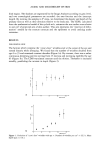
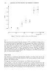
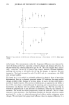
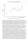
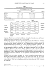
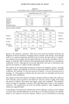
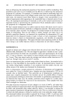
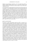
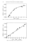
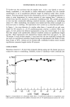
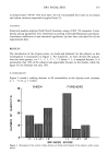
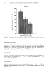
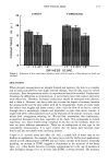
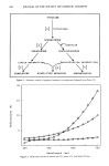
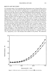
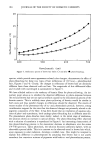
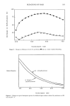
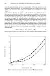
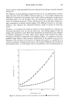
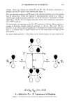
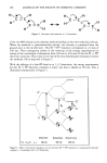
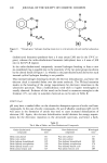
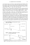
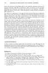
AGING AND MICRORELIEF OF SKIN 147 bital region. The shadows are segmented by the Image Analyzer according to grey level, and two stereological parameters are recorded: the area fraction and the intercept length. By rotating the samples in 9 ø steps, we determined the density and depth of the primary lines as well as their direction relative to the body axis. The CDSS, calculated from the mathematical model of the cycloid arch, estimates the true surface area relative to one cm 2 of projected area of skin surface. This area represents the "reservoir of defor- mation" needed by the stratum corneum and the epidermis to avoid cracking under extension. RESULTS PERI-ORBITAL SKIN The furrows which comprise the "crow's-foot" wrinkles start at the corner of the eye and extend linearly while diverging. We found that the number of wrinkles doubled from age 6 to 35 and remained constant therafter (Figure la). By contrast, there was a rather continuous deepening pattern starting from 25 microns and increasing eightfold by age 65 (Figure lb). The CDSS was almost constant until the thirties. Thereafter it increased steadily, paralleling the increase in depth (Figure 2). VlO a. a o age (years) + b o , elo • ,. •1o 40 eo =ge (yea•s) Figure 1. Evolution of "crow's foot" wrinkles with age. a. Number of wrinkles per cm 2 + S.E.b. Mean depth (I-tm + S.E.).
Purchased for the exclusive use of nofirst nolast (unknown)
From: SCC Media Library & Resource Center (library.scconline.org)