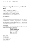
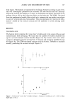
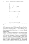
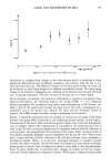
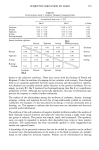
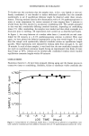
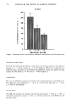
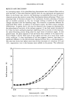
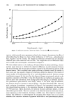
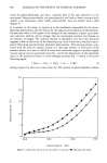
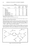
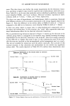
AGING AND MICRORELIEF OF SKIN 151 I .1 0 I .05. 1.00. 0 20 40 60 80 Figure 5. Leg: evolution of the CDSS versus age. age (years) informative to compare these changes to the volar forearm which is submitted to large amplitude deformations such as flexing, extension, and rotation. Like the leg, it is a relatively protected area. The different evolution of the skin relief in these two sites can be attributed to their being subjected to different mechanical stresses. The main aging change in the forearm's topography was a shifting of the primary lines towards the limb axis, increasing anisotropy. This also occurred in the leg, but to a lesser degree. On the forearm, accordingly, the reservoir of deformation, needed to accomodate these physical movements, was relatively high in the young (CDSS - 1.16). Repeated stresses throughout life necessarily bring about some reorientation of the furrows, no- tably a shift of the major lines towards the long axis of the limb, accompanied by a progressive loss of the second axis. Despite the latter and the decreased density of furrows, the CDSS actually increased owing to deepening of the furrows. Finally, it should be emphasized that the changes in orientation and depth of the major furrows with aging reflect alterations in the underlying dermal matrix. In this respect, deterioration of the elastic fiber network has greater impact than structural disorganiza- tion of collagen. Elastic fiber degeneration is noticeable at the ultrastructural level as early as age 30 when cavitations appear even in unexposed buttock skin (8). Likewise in protected skin, the subepidermal vertical skeins of fine elastic fibers, mainly bundles of microfibrils, gradually disappear and are completely lost by age 50 to 60. These events lead to laxity and loss of elastic rebound so that the skin becomes more vulnerable to mechanical stresses. When, as on the face, prolonged exposure to sunlight virtually destroys the elastic network, the skin becomes even looser and more prone to folding, being greatly in excess. Loss of collagen in elastotic tissue adds to this superabundant, loose, recoiless, flabby skin. In protected skin, changes in collagen are far less conspic- uous and of lesser importance compared to elastin. With aging, collagen becomes stiffer

152 JOURNAL OF THE SOCIETY OF COSMETIC CHEMISTS and less stretchable, exactly opposite to the observed alterations of the surface patterns. However, these dimensional SEM views of the collagen network show that the bundles become thicker, straighter, and more random (9). Moreover, probably owing to the diminution of the ground substance, the spaces between the bundles are narrower, resulting in tighter packing. We suggest that these changes probably determine the shift in the orientation of primary lines. In this regard, it has to be admitted that there is no good understanding of the dermal forces which determine the complex pattern of the microrelief. Langer's lines would reflect preferential orientation of the collagen bundles, but this has never been demonstrated convincingly. Langer found that round holes punched in ca- daver skin become elliptical, a phenomenon well known to surgeons. Old subjects were not included in his studies. We found (unpublished observations) that round holes in abdominal and buttock skin gaped and remained round, attributed mainly to deteriora- tion of the elastic fiber network. Our general thesis then is based on the understanding that the skin microrelief is quite susceptible to the effects of time (intrinsic aging). Changes in the microrelief can be enormously accelerated by actinic radiation (extrinsic photo-aging) which accelerates degradation of its elastic fibers (10). As a result, mechanical stresses can easily reorient the original patterns. REFERENCES (1) (2) (3) (4) (5) (6) (7) (8) (9) (10) P. Corneille, Stances. (XVIIth century). K. Hashimoto, New methods for surface ultrastructure, Internat. J. Dermatol., 13, 357-381 (1974). J. Wolf and S. Hanusova, Influence of age on skin relief in man. Folia Morphologica, 18, 262-280 (1970). R. M. Lavker, F. Kwong, and A.M. Kligman, Changes in skin surface patterns with age, J. Ger- ontol., 35(3), 348-354 (1986). P. Corcuff, J. de Rigal, J. L. Leveque, S. Makki, and P. Agache, Skin relief and aging, J. Soc. Cosmet. Chem., 34, 177-190 (1983). S. Makki, J. C. Barbenel, and P. Agache, A quantitative method for the assessment of the microtop- ography of human skin, Acta Dermatovener, 59, 285-291 (1979). P. Corcuff, F. Chatenay, and J. L. Leveque, A fully automated system to study skin surface patterns, Int. J. Cosmet. Sci., 6, 167-176 (1984). I. M. Braverman and E. Fonferko, Studies in cutaneous aging: I. The elastic fiber network, J. Invest. Dermatol., 78(5), 434-443 (1982). J. C. Smith and G. R. Finlayson, Dermal connective tissue alterations with age and chronic sun damage, J. Soc. Cosmet. Chem., 16, 525-535 (1965). A.M. Kligman, P. Zheng, and R. M. Lavker, The anatomy and pathogenesis of wrinkles, British J. Dermatol., 113, 37-42 (1985).
Purchased for the exclusive use of nofirst nolast (unknown)
From: SCC Media Library & Resource Center (library.scconline.org)