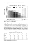
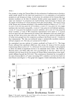
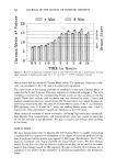
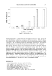
j. Soc. Cosmet. Chem., 41, 259-265 (September/October 1990) In vivo assessment of vesicant skin injury using a Minolta Chroma Meter ERNEST H. BRAUE, JR., MILLARD M. MERSHON, JOHN V. WADE, and MARTY R. LITCHFIELD, B•sic Assessment Br•nch, Drug Assessment Division, U.S. Army Medical Research Institute of Chemical DeJ•nse, Aberdeen Proving Ground, MD 21010-5425. Received September 4, 1990. Synopsis Current in vivo methods for evaluating the severity of vesicant skin lesions have relied on subjective visual scoring by trained observers. This report describes the development and evaluation of a novel method for the quantification of erythema caused by vesicant exposure to skin using the Minolta CR-200 Chroma Meter. Preliminary validation of this technique using a color chart with 19 different shades of red demon- strated a remarkable ability to distinguish between the different degrees of redness. Five replicate measure- ments on the 19 color zones yielded coefficients of variation (CV) in the 0.1% range. The experimental data demonstrated that the Chroma Meter's response was more sensitive than the human eye in differentiating small color differences. Preliminary validation of this technique has been demonstrated using euthymic hairless guinea pigs exposed to neat mustard (HD) vapor. The skin of 1 ! animals was exposed to HD vapor for either 4 or 8 minutes. Lesions were evaluated visually and with the Chroma Meter at times 0.5, 1, 1.5, 2, 3, 4, 5, 6, and 24 hours post-exposure. A time-response curve was established, and a significant correlation between the Chroma Meter response and the visual Draize scores was found. These studies have demonstrated that the Minolta Chroma Meter can be used to provide a reproducible, objective, and quanti- tative assessment of vesicant skin injury. INTRODUCTION The use of sulfur mustard and related vesicant compounds is well documented both in past and recent military conflicts. The identification of compounds that are effective in the treatment and prevention of injury caused by vesicants has been hampered by the lack of in vivo models for the quantitative and rapid assessment of prophylactic thera- peutic drugs. The Medical Chemical Defense Research Program would benefit greatly from an effective in vivo method for screening potential antivesicant compounds. Current in vivo methods for evaluating the severity ofvesicant skin lesions have relied on subjective visual scoring by trained observers. The human eye can serve quite well for qualitative visual evaluations of changes in skin caused by vesicant exposure, but the unaided human eye is extremely poor at extracting quantitative information. Another limitation is that observations may not be well correlated from one observer to another. The need for increased objectivity and quantitative accuracy in the scoring of chemically induced lesions has long been recognized however, visualization still persists as the 259

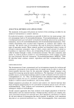
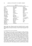
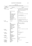
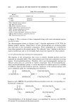
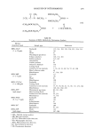
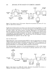
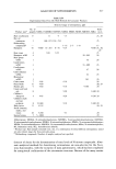
260 JOURNAL OF THE SOCIETY OF COSMETIC CHEMISTS major method of measurement in this field. There have been recent reports (1-12) that a reflectance color meter can be used for the quantitative evaluation of skin erythema. We have evaluated the ability of the Minolta CR-200 Chroma Meter (13) to provide reproducible and quantitative data for determining the degree of erythema caused by mustard (HD) vapor exposure. We used the euthymic hairless guinea pig as a novel animal model. The Chroma Meter used reflected light and read color in a three-dimen- sional format giving the brightness between black and white, the balance between red and green, and the relative amounts of yellow and blue. A preliminary study (14) from our laboratory evaluated the euthymic hairless guinea pig as an animal model for vesicant injury. The results demonstrated that this strain of guinea pig was considerably more sensitive than normal haired guinea pigs to the dermal injury produced by topical application of neat HD. In addition, the lack of hair in this strain greatly simplified the application of a vesicant agent and the subsequent evaluation of the dermal injury produced. This preliminary investigation was conducted in two phases. The first phase evaluated the Chroma Meter's response to small differences between shades of red color using a color chart. The second phase compared the Chroma Meter's response to visual Draize scores of the erythema that developed on the skin of euthymic hairless guinea pig exposed to HD vapor. METHODS A Minolta Chroma Meter model CR-200 (13) was used to make all measurements. This instrument is commercially available and includes a small hand-held measuring head connected to a portable data-processing unit by a flexible cord. The optical system of the measuring head illuminated the sample using diffuse light produced by a pulsed xenon arc lamp with a viewing angle of 0 ø. The area measured was a circle 8 mm in diameter. A total of six silicon photocells were used by a double-beam feedback system to ensure accurate and consistent measurements. Three of the photocells monitored the output of the pulsed xenon arc lamp the other three photocells measured the light reflected by the surface of the sample. The detected signal was converted into three coordinates (L*, a*, and b*) of a three-dimensional color system recommended by CIE (Commission Internationale de I'Eclairage) (15). The coordinate L* represented levels of brightness between white (+ 100) and black (- 100). The a* represented the relative chromaticity between red (+ 60) and green (- 60). The b* coordinate represented the relative amounts of yellow ( + 60) and blue ( - 60). A complete discussion on CIE color systems may be found in the book by Hunt (16). PHASE 1 A color chart was prepared which had 19 zones ranging in color from pale pink to bright red. Five replicate measurements were recorded for each zone and the data ana- lyzed. PHASE 2 The skin of hairless guinea pigs was exposed to HD vapor for either 4 or 8 minutes by a vapor cup exposure technique developed in this laboratory. Guinea pigs were anesthe-
Purchased for the exclusive use of nofirst nolast (unknown)
From: SCC Media Library & Resource Center (library.scconline.org)