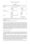
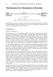
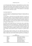
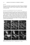
110 JOURNAL OF THE SOCIETY OF COSMETIC CHEMISTS the method, dimensional stability of the negative replica, and ability of the method to reproduce fine, delicate structures were addressed. The method is applied to the deter- mination of the fate of an experimental polymer contained in a facial lotion applied to the forehead. EXPERIMENTAL REPLICATION The in vivo skin replication method involved a two-step process, as illustrated in Figure 1. The first step consisted of making a negative impression of the surface of the skin with a rubbery vinyl polysiloxane material, Reprosil (Dentsply International, York, PA). The second step consisted of producing a positive impression from the Reprosil negative using a low-viscosity epoxy resin, Spurr's embedding medium (Electron Microscopy Sciences, Fort Washington, PA). To make a negative impression of the skin, the following procedure was used. For experimental procedures utilizing replication methods to study skin surface topography, it was imperative that the identical site on the skin be replicated each time. Ink from a ball-point pen was easily transferred to the Reprosil impression material from the skin. A mark (e.g., an arrow) was made on the surface of the skin under study with a ball-point pen. The mark was refreshed before each replica was made. For long-term studies, participants were asked to refresh the mark each day. The Reprosil was prepared as described in the instructions contained in the kit. The mixed material was applied to the area of skin under study, as a thin film, approximately 1 to 2-mm thick. A one-inch-diameter circular piece of 3.2-mm-thickness Teflon polymer sheet was next applied to smooth out the replica and provide a flat surface necessary for subsequent REPLICATION PROCESS SKIN REPROSIL NEGATIVE EPOXY • POSITIVE Figure 1. Schematic of skin replication process.

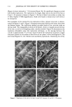
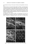
REPLICATION OF SKIN 111 steps. The material was allowed to cure for a minimum of eight minutes from the start of the mix. (Variations in temperature will affect the work and set times. Accordingly, the replica was not to be removed from the skin until it was firm, resilient, and non-tacky.) The replicas were stored in Petri dishes to protect the surface. It is suggested to delay making the epoxy impression for 60 minutes. To make a positive impression for the negative replica, Spurr's low-viscosity embedding medium was prepared according to directions contained in the kit. The positive replica was generated using a circular mold that was 2-cm in diameter and 0.5-cm deep. The mold was filled with the epoxy resin using a disposable pipet. The negative impression was precut to fit in the mold. A drop of resin was applied to the replica and carefully spread to wet the surface. The negative was gently placed into the filled mold, surface- down, onto the epoxy. (The Reprosil will float and can be cured in this manner.) The molds were placed in a large glass Petri dish and cured at 65øC in an oven in a fume hood for a minimum of eight hours. WARNING: Do not use any containers made of polystyrene with the Spurr's embedding medium as the polystyrene is incompatible (i.e., the Spurifs medium will dissolve the polystyrene). After curing, the samples were removed form the molds upon cooling to room tem- perature. The positive impressions were trimmed to remove excess epoxy material by scoring the undersurface and cutting from the opposite side. The Reprosil was carefully peeled away from the cleaned epoxy replica. The positive impression was mounted on a one-inch, aluminum scanning electron microscope (SEM) stub with double-sided ad- hesive. The specimen was then sputter-coated with approximately 25 nm of Au/Pd. iNSTRUMENTATION The positive impressions were examined with a Hitachi S-520 SEM operating at l0 KV, 15 mm working distance, and a 15 ø tilt towards the detector. DIMENSIONAL STABILITY OF THE REPROSiL NEGATIVE IMPRESSION An impression of an area of approximately 30 mm 2 on the inner left forearm was made as described above. A positive impression was produced from the same Reprosil mold on days 0, 77, and 81. EFFECT OF REPLICATION ON THE SURFACE OF THE SKIN Impressions of an area of approximately 30 mm 2 on the inner left forearm were made in series at 30-minute intervals. Reprosil impressions of the sole of the foot were made at 0 and 30 minutes. REPLICATION OF FINE, DELICATE STRUCTURES An impression of an area of the forearm that contained several hair follicles was made as described above. In addition, replicas of the leaves of two plants, velvet leaf and coleus, were also made.
Purchased for the exclusive use of nofirst nolast (unknown)
From: SCC Media Library & Resource Center (library.scconline.org)