INFLUENCE OF LIPOSOMAL ENCAPSULATION 121 METHODS Preparation and characterization of liposomes Preparation. Liposomes were prepared by a combination of Bangham's film hydration method (23) and the extrusion technique described by Nayar et al. (24). First, t-RA, trace amounts of [3H]t-RA, and the lipids in chloroform solutions were mixed together in the appropriate ratio and 1% of ot-tocopherol (based on total lipid weight) was added as an antioxidant. For PC/t-RA liposomes, the ratio was 16 •mol PC, 4 •mol t-RA, and 50 •Ci [3H]t-RA for the low-dose studies, and 16 •mol PC, 4 •mol t-RA, and 1.2 •Ci [3H]t-RA for the high-dose studies for PC/PE/OA/CHEMS/t-RA liposomes, it was 4 •mol PC, 4 •mol PE, 2 •mol OA, 10 •mol CHEMS, 4 •mol t-RA, and 50 •Ci [3H]t-RA for the low-dose studies, and 4 btmol PC, 4 •mol PE, 2 I-tmol OA, 10 •mol CHEMS, 4 •mol t-RA, and 1.2 •Ci [3H]t-RA for the high-dose studies. The lipid- drug mixture was deposited as a thin film in a round-bottom flask by roto-evaporating the chloroform (Rotavapor RE 111 ©, Buchi, Switzerland) under a gentle nitrogen stream. Vacuum was applied for one hour to ensure total removal of trace solvents. The film was then hydrated at 40øC for one hour with HEPES buffer (20 mM HEPES, 150 mM NaC1, 0.1 mM EDTA). HEPES buffer, pH 7.4, was used for the PC liposomes and HEPES buffer, pH 8.0, for the PC/PE/OA]CHEMS liposomes. After hydration was complete, the preparation was sonicareal for 30 rain (Ultramet III sonic cleaner, Buehler Ltd., Evanston, IL) and five freeze-thaw cycles were performed (5 min, dry ice/40øC waterbath). When necessary, the pH of the PC/PE/OA/CHEMS dispersion was adjusted back to 8.0 with 1 N NaOH. The resulting large multilamellar vesicle dispersion was then transferred into a stainless steel extrusion device (The Extruder TM, Lipex Biomembranes, Vancouver, BC), and unilamellar liposomes were generated by forcing the preparation through two stacked polycarbonate filters (Nuclepore Corp., Pleasanton, CA) of defined pore size (400 nm, 200 nm, 100 nm, or 50 nm in diameter). Nitrogen pressures up to 2800 kPa (400 psi) were used, and all extrusions (ten passes for each preparation) were performed at room temperature. After extrusion was complete, the volume of preparation was adjusted with buffer so that the final suspension contained 0.05% w:v [3H]t-RA. For the low-dose studies, [3H]t-RA specific activity in the final preparation was 12.5 •Ci/•mol, and its radiochemical concentration was 21 •Ci/ml. For the high-dose studies, [3H]t-RA spe- cific activity was 0.3 •Ci/•mol, and its radiochemical concentration was 0.5 •Ci/ml. The final lipid concentration was about 6 mg/ml in both cases. The liposome prepara- tions were stored at 4øC under nitrogen before use. Size determination. We determined the particle size distribution of the extruded liposomal systems by quasi-elastic light scattering (QELS) using a Malvern 4700c submicron particle size analyzer (Malvern Instruments, Southborough, MA) equipped with a 60 mW helium-neon laser at an excitation wavelength of 633 nm. The temperature was set at 25øC and measurements were taken at 90 degrees. In vitro skin permeation studies Preparation of control formulations. For the low-dose studies, we prepared an alcoholic [3H]t-RA formulation (50 •Ci/ml, 12.5 •Ci/•mol) by spiking a 0.05% ethanolic solution of t-RA with trace amounts of radioactive t-RA. Alcohol has been shown to be an efficient delivery vehicle for t-RA (25) and is used in commercial topical preparations

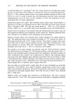
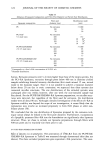
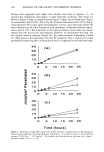
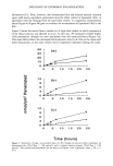
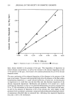
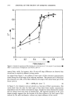
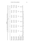
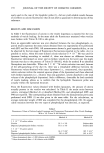
122 JOURNAL OF THE SOCIETY OF COSMETIC CHEMISTS of t-RA like Retin-A TM and Aberel © (16). The control vehicle for the high-dose studies and one set of low-dose studies (Figure 3) consisted of a mixture of transcutol and water, 68:32 v:v. This cosolvent system yields a very nearly saturated solution of t-RA at the 0.05% w:v level. In order to ensure that this formulation did yield nearly maximum thermodynamic activity of t-RA, we also included a 0.10% t-RA suspension in trans- cutol/water 68:32 in some experiments. Preparation of human skin samples. Split-thickness human cadaver skin, dermatomed to a thickness of 250 •tm, was obtained from the Ohio Valley Skin 8: Tissue Center (Vernon Place, Cincinnati, OH). The skin was stored frozen at -80øC in a 10% glycerol solution. Before use, it was rapidly thawed and thoroughly rinsed with distilled water. Isolated sheets of epidermis were prepared from the dermatomed skin samples using the heat separation method (2-min immersion in 60øC water) (26). Isolated epidermal sheets were mounted on the diffusion cells immediately after preparation. Stratum corneum sheets were prepared from isolated epidermis by trypsin digestion (26). The epidermal sheets were incubated overnight at 37øC, dermal side down, on filter paper saturated with 0.01% trypsin solution. The stratum corneum sheets were then rinsed with a 0.007 % trypsin inhibitor solution, followed by distilled water. They were mounted on 20-mm drain discs (Nuclepore Corp., Pleasanton, CA) for easy handling, and stored dry at -20øC in a dessicator until needed. The integrity of all tissue samples was assessed using the 3H20 permeation method described by Franz and Lehman (27). The samples yielding a water flux greater than 1.2 mg/cm 2 were discarded unless otherwise noted. For comparison, the barrier integrity of some dermatomed skin samples was compromised by adhesive tape-stripping (about nine strips with 3M cellophane tape). Example results from that test are shown in Table I. Dermatomed skin, isolated epidermis, and isolated stratum corneum yielded equiv- alent water permeation rates, comparable to the values found by Franz and Lehman (27). Tape-stripped skin, as expected, gave a higher water flux. Care was taken to use skin from a single donor for all types of samples in a particular experiment. Nevertheless, since quality variability among packs of skin from a single donor was sometimes experienced, the water flux geometric mean of each set of samples is presented in the figure legends below. Note that the data in Table I were obtained from a single pack. Diffusion studies. All samples were mounted on modified Franz cells with a nominal surface area of 0.79 cm 2 (28). The receptor solution was Dulbecco's phosphate-buffered Table I Water Permeability of Different Skin Preparations From One Donor as Measured by Franz and Lehman's Method 3H20 penetration, mg/cm 2 Preparations Geometric mean SE n Dermatomed skin 0.27 0.04 7 Isolated epidermis 0.27 0.04 7 Isolated stratum corneum 0.21 0.07 10 Tape-stripped skin 1.34 0.22 7
Purchased for the exclusive use of nofirst nolast (unknown)
From: SCC Media Library & Resource Center (library.scconline.org)