168 JOURNAL OF THE SOCIETY OF COSMETIC CHEMISTS The aim of this work is to reproduce the same type of experiments with a hydrophilic fluorescent model molecule (calcein) and to compare the new results with those obtained with DPH. The choice of calcein is related to its self-quenching properties that make this substance a useful tool in liposome investigations (3). By means of this study we intend also to verify if empty liposomes, dispersed in a calcein solution, are capable of incorporating a certain amount of this substance. This last point acquires a fundamental importance if we consider that in the formulation of cosmetic preparations containing liposomes, hydrophilic substances can be present (e.g. preser- vatives, fragrances, coloring agents, etc.) and that their properties can be modified by incorporation in the vesicle structure furthermore, since liposomes have been claimed to enhance skin permeation, these substances could become, in the presence of lipo- somes, the cause of allergic or toxic effects. Studied liposomes were obtained by sonication, although other techniques (e.g. reverse- phase evaporation) may yield much higher trapping efficiency for hydrophilic substances (4). Sonication was chosen for this investigation because it is a procedure commonly used in cosmetics. MATERIALS 99%-pure L-ot-phosphatidylcholine from egg yolk (Sigma, type XI-E, chloroform so- lution, 100 mg/ml) and 90%-pure enriched soya phosphatidylcholine (Phospholipon 90, Nattermann Phospholipids GmbH) were used for vesicle preparation. Crystalline calcein was purchased from Sigma. HEPES pH 7.5 buffer solutions (10- 3 M), obtained with freshly distilled and aleaerated water, were used. Cholesterol, Triton X- 100, and all other products used for the present investigation were of analytical grade. All solvents were tested for fluorescence at the wavelength of interest for our studies. As in the previous work (1), fluorescence (Ex. 490 nm/Em. 520 nm) and turbidity (600/600 nm) measurements were carried out with a Perkin Elmer LS5 spectrofluorom- eter. Sonication was performed with a Soniprep 150 apparatus (MSE, Crowley). Lipo- some dimensions were evaluated with a Malvern Autosizer II. The Phospholipids B test kit (Wako Chemicals GmbH) was used for quantitative determinations of these sub- stances. Calcein solutions of two different concentrations (5 x 10⁻⁵ and 5 x 10 -2 M) were used for the present investigation these were obtained by dissolving an exactly weighed amount of the fluorescent probe in the minimum volume of a 1 M NaOH solution. The final calcein concentrations were then reached by addition of the HEPES solution (3). The pH (7.5) was checked each time by means of a potentiometer. The 5 x 10⁻⁵ M calcein solution was chosen because it had a concentration similar to that obtainable with DPH (1), while the more concentrated dye solution was used to constrain the hydro- philic probe within the liposomal structure. METHODS As in the previous study, vesicles containing the probe were prepared according to two different protocols.

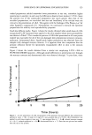
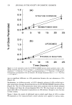
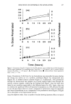
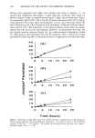
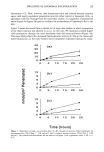
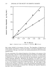
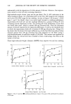
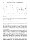
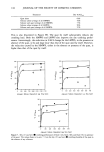
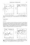
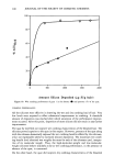
LIPOSOMES IN COSMETICS 169 METHOD A (SONICATION IN THE PRESENCE OF CALCEIN) The appropriate amount of phospholipid (80 mg of P90 or 800 }•1 of EPC solution) and 5.6 mg of cholesterol were completely dissolved in 4-5 ml of methanol. The solvent was vacuum-evaporated to form a thin film of lipids and additive inside the vessel. 2.5 ML of one of the two calcein solutions in the HEPES buffer were added, and the mixture was gently shaken for 1 h and sonicated, under a nitrogen stream, for 40 min (eight times for 5 min). The temperature was maintained at 15-20øC by means of a water bath. The liposome dispersion was finally diluted 1:1 with HEPES buffer. METHOD B (SONICATION IN THE ABSENCE OF CALCEIN) Liposomes were prepared according to the same procedure described above, but no marker was added until the final dilution. This 1:1 dilution, at the end of the vesicle preparation, was performed with the same HEPES solution of calcein used in method A. Unmarked liposomes were kept in the dark overnight with one of the fluorophore solutions. Longer times did not significantly increase the amount of absorbed calcein. It has been pointed out (5) that sonication of phospholipid dispersions leads mainly to small unilamellar vesicles. In our studies, liposome sizes ranged from 25 to 40 nm. The reproducibility of the different preparations was checked by turbidity measurements. Liposome separation from "free" calcein and "free" phospholipids was performed on 1.0 ml samples with Sephadex G75. Columns were eluted with HEPES, and all the vesicles were collected (liposomes were eluted with the void volume, and their presence was checked by a turbidity test) to reach a final volume of 5 mi. The Phospholipids B test was performed before and after the passage through the columns in order to verify the percentage of aggregated form with respect to the total amount used. The results indicated that over 95% of the initial amount of phospholipids was always recovered in the form of liposomes. All final preparations containing the vesicles were tested for turbidity. The reproducibility of these last measurements (fluc- tuations of +-3.5%), performed on the different preparations, indicated that the average dimensions and concentration of liposomes were to be considered as constant (6). Calcein fluorescence was initially determined on intact purified liposomes in order to verify once more the reproducibility among the various preparations of the same kind. For the determination of the total amount _of_ calcein present in the vesicles, they were broken by addition of a 1% Triton X-100_HEPES solution. This surfactant, at appro- priate concentrations, is capable of breaking down the liposomal structure (1,7), and at the same time it allows a correct quantitative evaluation of calcein because its effect on --2 the fluorescence behavior of the marker was recently studied (8,9). When the 5 X 10 M calcein solution was used, in order to reach measurable fluorescence values and because of the remarkably different amount of calcein in the liposomes according to the loading technique, a further 1/200 or 1/20 dilution was carried out for liposomes obtained with methods A and B, respectively L By these dilutions it was also possible to have solutions where fluorescence was linearly dependent on calcein concentration. In all cases, for an appropriate comparison, dispersions of intact liposomes were diluted ac- cordingly. Calcein concentrations were calculated from appropriate calibration curves of the marker in HEPES and Triton X-100 (9). Liposome rupture with methanol, previ-
Purchased for the exclusive use of nofirst nolast (unknown)
From: SCC Media Library & Resource Center (library.scconline.org)