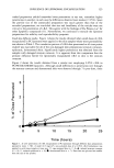
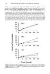
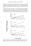
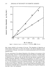
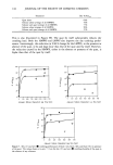
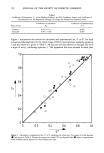
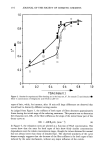
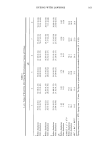
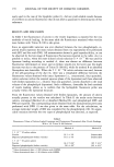
170 JOURNAL OF THE SOCIETY OF COSMETIC CHEMISTS ously used in the case of the lipophilic probe (1), did not yield reliable results because of its effects on calcein fluorescence that do not allow a quantitative determination of this substance. RESULTS AND DISCUSSION In Table I the fluorescence of calcein in the vesicle dispersion is reported for the two methods of vesicle loading. In the same table the fluorescence measured when vesicles were broken with Triton X-100 is also given. Since no appreciable variation was ever observed between the two phospholipids, re- ported results represent the mean values obtained from ten experiments (five performed with EPC and five with P90). All measurements showed a good reproducibility, as can be observed by the low range of fluorescence fluctuations reported in the table. As it is possible to notice, when the more diluted calcein solution (5 x 10⁻⁵ M) was used for liposome loading according to method A, there was almost no difference between fluorescence determined on intact and on broken vesicles [in the latter case the slight decrease was due to the presence of Triton X-100 (9)], while for method B no absorbed fluorophore was detectable. When the 5 X 10⁻² M calcein solution was used, because of the self-quenching of the dye (3), there was a remarkable difference between the fluorescence values obtained with intact liposomes (i.e., concentrated, thus quenched, calcein solution within the internal aqueous phase of the liposomes) and those observed with broken liposomes (i. e., diluted, thus non-quenched, calcein dissolved in the total volume of the phospholipid dispersion). Such a difference, detectable for both methods of vesicle loading, allows us to confirm that the hydrophilic fluorescent probe was actually within the liposomal structure. From the fluorescence values obtained with broken liposomes, the amount of calcein actually present in the vesicles was calculated. In Table II the molar ratios between calcein, entrapped (Method A) or absorbed (Method B), and phospholipid (EPC and P90) are reported. The corresponding values obtained from the measurements previously performed with DPH (1) are also given in the same table. For the calculations, an average molecular weight of 800 was considered for the phospholipids (10). No appre- ciable variation between the two types of phospholipids was detected, as expected. Table I Fluorescence Values of Calcein-Loaded Liposomes and Fluorescence Determined After Vesicle Breakage With a 1% Triton X-100 Solution Method A Method B 5 X 10 -5 M calcein solution Vesicles Dispersed phospholipids (broken vesicles) 5 x 10 - 2 M calcein solution Vesicles Dispersed phospholipids (broken vesicles) 39.5 + 5% Undetectable 35.0 + 5% Undetectable 16.4 --+ 6% • 185 --+ 4% 2 150 --- 4% • 365 ñ 4% 2 Values determined after a 1:200 dilution with HEPES. Values determined after a 1:20 dilution with HEPES.

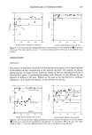
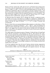
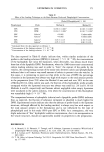
LIPOSOMES IN COSMETICS 171 Table II Effect of the Loading Technique on the Ratio Between Probe and Phospholipid Concentrations Phospholipid Probe [Probe]/[Phospholipids] X 100 Method A Method B EPC DPH P90 DPH EPC Calcein 2 P90 Calcein 2 EPC Calcein 3 P90 Calcein 3 0.036 - 0.003 • 0.034 + 0.003 • 0.002 0.002 3.03 - 0.15 2.95 --- 0.15 0.018 + 0.003 • 0.016 -+ 0.0031 Undetectable Undetectable 0.78 -+ 0.05 0.80 --- 0.05 Calculated from the data reported in reference 1. Concentration of the loading solution: 5 X 10⁻⁵ M. Concentration of the loading solution: 5 X 10⁻² M. The data reported in Table II clearly indicate that, within similar molarities of the probes in the loading solutions ([DPH] --• [calcein] = 5 X 10⁻⁵ M), the concentration of the hydrophilic dye in/on the liposomes, when detectable, was always much lower than that of the lipophilic DPH. Furthermore, when the 1000-times-more-concentrated calcein loading solution was used in order to "force" the capture of the probe by the vesicles, the corresponding increase of the molar ratio between calcein and phospholipids indicated that in all cases a very small amount of calcein is present in/on the vesicles. In this sense, it is interesting to point out that while in the case of DPH the percentage of marker in the liposomes was always very high with respect to the total amount present in the preparation [over 70% when the Method A was used and over 50% in the case of Method B (1)], when calcein was used, the fraction of this hydrophilic probe actually entrapped within the liposomal structure was always very small (ca. 2% and 0.1% for Methods A and B, respectively) and became almost negligible when empty liposomes were incubated in the calcein solution, even when the concentration of this fiuorophore was remarkably high (5 x 10⁻² M). The comparison between a hydrophilic (calcein) and a lipophilic probe (DPH) indicates that, as expected, the fraction of substance entrapped or absorbed is much higher for DPH. Experimental results indicate also that the amount of probe found in the liposome structure, although affected by the loading method, is always very low with respect to the total amount present in the formulation and is not influenced by the type of phospholipid used in this study (99%-pure EPC or P90). As a consequence, the very slight reduction of"free" hydrophilic additive concentration, due to its incorporation in the vesicle structure, should not appreciably modify its properties in the preparation. REFERENCES (1) A. Memoli, L. G. Palermiti, V. Travagli, and F. Alhaique, Liposomes in cosmetics: Which kind of phospholipid? Which loading method?, J. Soc. Cosm. Chem., 44, 123-128 (1993). (2) B. J. Litman and Y. Barenholz, Fluorescent probe: Diphenyhexatriene, Methods Enzymol., 81, 678- 685 (1982). (3) T. M. Allen, in Liposome Technology, Vol. III, G. Gregoriadis, Ed. (CRC Press, Boca Raton Fl., 1984), pp. 177-182.
Purchased for the exclusive use of nofirst nolast (unknown)
From: SCC Media Library & Resource Center (library.scconline.org)