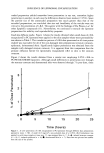
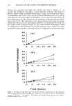
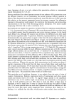
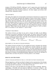
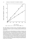
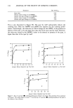
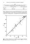
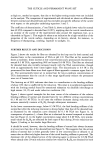
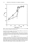
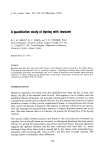
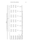
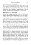
INFLUENCE OF LIPOSOMAL ENCAPSULATION 125 truded preparation yielded somewhat lower penetration in one test, somewhat higher penetration in another in each case the differences observed were modest (25%). Since the particle size of the unextruded preparation was much greater than that of the extruded preparations, we concluded that size and lamellarity of the vesicles were not critical to the penetration of t-RA. This agrees with the findings of Du Piessis et al. for other lipophilic compounds (11). Nevertheless, we continued to extrude the liposome preparations for stability and reproducibility purposes. Small-dose diffusion studies. Figure 1 shows the results obtained when small doses of t-RA encapsulated in PC liposomes were applied to the skin samples whose water permeability was shown in Table I. The cumulative percent of t-RA dose penetrated at all time points studied was equivalent for all of the non-damaged skin preparations (stratum corneum, epidermis, dermatomed skin). Significantly higher penetration was obtained from the samples with damaged stratum corneum. It is apparent from this comparison that the primary diffusion barrier for liposomally encapsulated t-RA in skin is the stratum corneum. Figure 2 shows the results obtained from a similar test employing 0.05% t-RA in PC/PE/OA/CHEMS liposomes. Although small differences in penetration rate through the stratum corneum and dermatomed skin were observed through 7 h post-dose, there 0 L • 0 5 10 15 20 25 Time (hours) Figure 1. In vitro penetration of t-RA encapsulated in PC liposomes through different skin preparations (geometric mean -+ SE). A small (4.8 mg/cm2), non-occluded dose of a 0.05% t-RA formulation was applied to each tissue sample at time zero. O, tape-stripped skin (n = 4) &, isolated stratum corneum (n = 8) /•, isolated epidermis (n = 6) O, dermatomed skin (n = 7). Error bars not pictured are smaller than the size of the symbol.

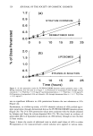
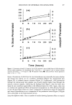
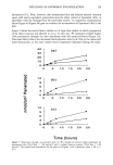
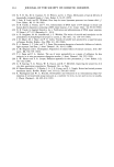
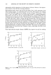
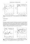
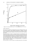
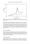
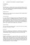
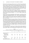
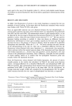
126 JOURNAL OF THE SOCIETY OF COSMETIC CHEMISTS o 1.2 [ (a) O9 O6 O3 • DERMATOMED SKIN 00 ' ' ' ' 0 5 10 15 20 25 2.0 (b) I 5 lO 0 5 • , ETHAN?LIC SOL, UTION O0 ' 0 5 10 15 2o 25 Time (hours) Figure 2. In vitro penetration results for PC/PE/OA/CHEMS liposome system (geometric mean + SE). Dosing conditions were the same as in Figure 1. (a) Penetration of encapsulated t-RA through stratum corneum (n = 4, 3H20 flux = 0.35 mg/cm 2) and dermatomed skin (n = 7, 3H20 flux = 0.34 mg/cm2) (b) Penetration of encapsulated t-RA and ethanolic t-RA through dermatomed skin (n = 6-7, 3H20 flUX = 0.33 mg/cm2). was no significant difference in t-RA penetration between the two substrates at 24 h (Figure 2a). Furthermore, in a follow-up study, a 0.05% ethanolic solution of t-RA yielded equiv- alent penetration through dermatomed skin as the PC/PE/OA/CHEMS liposome system (Figure 2b). The 24 h t-RA penetration values were consistent with the results obtained by Lehman and Franz for solvent-deposited RA (25). We thus found no evidence of appreciable effects of liposomal encapsulation on t-RA delivery through or into the skin in these studies. Figure 3 shows the results of additional tests in which small doses of t-RA in either PC-liposomes or the transcutol/water control solution were applied to various mem-
Purchased for the exclusive use of nofirst nolast (unknown)
From: SCC Media Library & Resource Center (library.scconline.org)