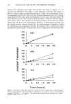
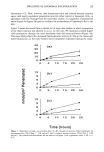
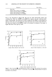
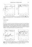
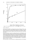
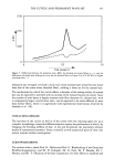
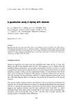
DYEING WITH LAWSONE 165 neutralization mechanism suggested above permits absorption of species II onto the keratin of hair at low pH values, giving a "reddish-orange" hue, but does not explain why the hue is more yellow when the hair is dyed at high pH values. Forestier (9) postulated complexation involving cystine sulfur or an amine function in keratin and the 3-carbon of lawsone, exemplified by (IV) and (V). O O ( S-Keratin ( N-Keratin However, attack of keto-carbon in lawsone, to give a Schiff's base as exemplified by Eq. 3, appears more likely: C9H502' - C -- O q- H2N - Keratin = C9H502' - C = N - Keratin + H20 (Lawsone) (Free NH 2 in (Schiff's base) (Eq. 3) keratin) A similar reaction involving the hydrosulfide groups of keratin could also occur. REFERENCES (1) K. C. James, S. p. Spanoudi, and T. D. Turner, The absorption of lawsone and henna by bleached wool felt,.]. Soc. Cosmet. Chem., 37, 359-367 (1986). (2) L. I. Savraanskii and A. T. Pilipanko, Tautomerism and structure of anion and cation forms of hy- droxynaphthoquinones, Dopov. Akad. Nuak. Ukr., RSR Ser. B, 33, 829-833 (1971). Through Chem. Abs., 76, 13658q. (3) H. H. Hodgson and H. S. Turner, Colour and constitution. Part VII--Some observations on the structures of the mono- and di-nitronaphthylamines based on their visual colours. The probable structure of B-napthaquinone, J. Soc. Dyers Colorists, 59, 218-220 (1943). (4) N. N. Shapet'ko and D. N. Shigorin, Proton NMR in the O-H... O intramolecular H-bond in quinoid structure, Zh. Strukt. Khim., 8, 538-540 (1967). Through Chem. Abs., 67, 103798d. (5) B. I. Amro, Dyeing With Henna and Related Materials, PhD Thesis, University of Wales, 1989. (6) M.-L. Josien, N. Fuson, J.-M. Lebas, and T. M. Gregory, An infrared spectroscopic study of the carbonyl stretching frequency in a group of ortho and para quinones, J. Chem. Phys., 21, 331-340 (1953). (7) L. F. Fieser and M. Fieser, Advanced Organic Chemistry (Chapman and Hall, London, 1961), p. 846. (8) C. R. Robbins, Chemical and Physical Behaviour of Human Hair (Van Nostrand Reinhold, New York, 1979), p. 96. (9) J.P. Forestier, Henne: Absorption de la lawsone par le cheveu, Int. J. Cosmet. Sci., 4, 153-174 (1982).

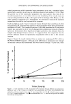
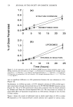
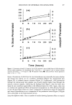
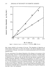
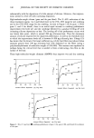
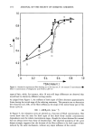
j. Soc. Cosmet. Chem., 45, 167-172 (May/June 1994) Liposomes in cosmetics. II. Entrapment of a hydrophilic probe ADRIANA MEMOLI, LUISA G. PALERMITI, VALTER TRAVAGLI, and FRANCO ALHAIQUE, Dipartimento di Studi di Chimica e Tecnologia delle Sostanze Biologicamente Attive, Universita di Roma (La Sapienza) (A.M., L.G.P.), Dipartimento Farmaco Chimico Tecnologico, Universita di Siena (V. T. ), and Dipartimento Farmaco Chimico Tecnologico, Universita di Cagliari (F.A. ), Italy. Received January 6, 1994. Synopsis Phospholipids of different origin (egg and soya) and purity were used to prepare liposomes by sonication. Loading of these vesicles with a fluorescent hydrophilic model molecule (calcein) was carried out by means of two different methods. No appreciable differences were observed in the loading capacities of liposomes prepared with the two products. Obtained results indicated that the fraction of incorporated or absorbed calcein was always very small with respect to the total amount of this substance used for the preparation of the vesicles furthermore, the quantity of hydrophilic probe in the vesicle structure was remarkably affected by the loading method. The results of this investigation allowed us to consider the possible incorporation, in/on the vesicles, of additives that can be present in a formulation containing liposomes. INTRODUCTION It is well known that liposome entrapment capacity depends on several factors, such as the type of phospholipid, the physicochemical properties of the substance that must be loaded, and the technique used for the preparation of the vesicles. In a previous paper (1), in order to compare the behavior of a 99%-pure egg phosphati- dylcholine (EPC) with that of a mdch less expensive, but also less pure, vegetable phospholipid (P90), a lipophilic fluorescent probe (1,6-diphenyl-l,3,5-hexatriene DPH) (2), localized within the lipid bilayers, was chosen for our experiments. The results obtained (1) indicated that no remarkable differences were observed in either the stability in regard to surfactant-induced breakage or the loading capacity of liposomes respectively prepared with EPC or P90. It was also shown that the amount of lipophilic probe in the liposome structure was affected by the loading method. 167
Purchased for the exclusive use of nofirst nolast (unknown)
From: SCC Media Library & Resource Center (library.scconline.org)