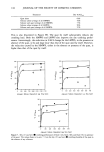
LIPOSOMES IN COSMETICS 169 METHOD A (SONICATION IN THE PRESENCE OF CALCEIN) The appropriate amount of phospholipid (80 mg of P90 or 800 }•1 of EPC solution) and 5.6 mg of cholesterol were completely dissolved in 4-5 ml of methanol. The solvent was vacuum-evaporated to form a thin film of lipids and additive inside the vessel. 2.5 ML of one of the two calcein solutions in the HEPES buffer were added, and the mixture was gently shaken for 1 h and sonicated, under a nitrogen stream, for 40 min (eight times for 5 min). The temperature was maintained at 15-20øC by means of a water bath. The liposome dispersion was finally diluted 1:1 with HEPES buffer. METHOD B (SONICATION IN THE ABSENCE OF CALCEIN) Liposomes were prepared according to the same procedure described above, but no marker was added until the final dilution. This 1:1 dilution, at the end of the vesicle preparation, was performed with the same HEPES solution of calcein used in method A. Unmarked liposomes were kept in the dark overnight with one of the fluorophore solutions. Longer times did not significantly increase the amount of absorbed calcein. It has been pointed out (5) that sonication of phospholipid dispersions leads mainly to small unilamellar vesicles. In our studies, liposome sizes ranged from 25 to 40 nm. The reproducibility of the different preparations was checked by turbidity measurements. Liposome separation from "free" calcein and "free" phospholipids was performed on 1.0 ml samples with Sephadex G75. Columns were eluted with HEPES, and all the vesicles were collected (liposomes were eluted with the void volume, and their presence was checked by a turbidity test) to reach a final volume of 5 mi. The Phospholipids B test was performed before and after the passage through the columns in order to verify the percentage of aggregated form with respect to the total amount used. The results indicated that over 95% of the initial amount of phospholipids was always recovered in the form of liposomes. All final preparations containing the vesicles were tested for turbidity. The reproducibility of these last measurements (fluc- tuations of +-3.5%), performed on the different preparations, indicated that the average dimensions and concentration of liposomes were to be considered as constant (6). Calcein fluorescence was initially determined on intact purified liposomes in order to verify once more the reproducibility among the various preparations of the same kind. For the determination of the total amount _of_ calcein present in the vesicles, they were broken by addition of a 1% Triton X-100_HEPES solution. This surfactant, at appro- priate concentrations, is capable of breaking down the liposomal structure (1,7), and at the same time it allows a correct quantitative evaluation of calcein because its effect on --2 the fluorescence behavior of the marker was recently studied (8,9). When the 5 X 10 M calcein solution was used, in order to reach measurable fluorescence values and because of the remarkably different amount of calcein in the liposomes according to the loading technique, a further 1/200 or 1/20 dilution was carried out for liposomes obtained with methods A and B, respectively L By these dilutions it was also possible to have solutions where fluorescence was linearly dependent on calcein concentration. In all cases, for an appropriate comparison, dispersions of intact liposomes were diluted ac- cordingly. Calcein concentrations were calculated from appropriate calibration curves of the marker in HEPES and Triton X-100 (9). Liposome rupture with methanol, previ-

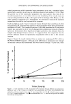
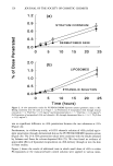
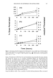
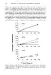
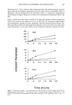
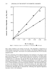
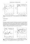
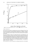
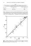
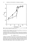
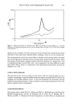
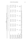
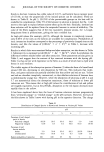
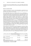
170 JOURNAL OF THE SOCIETY OF COSMETIC CHEMISTS ously used in the case of the lipophilic probe (1), did not yield reliable results because of its effects on calcein fluorescence that do not allow a quantitative determination of this substance. RESULTS AND DISCUSSION In Table I the fluorescence of calcein in the vesicle dispersion is reported for the two methods of vesicle loading. In the same table the fluorescence measured when vesicles were broken with Triton X-100 is also given. Since no appreciable variation was ever observed between the two phospholipids, re- ported results represent the mean values obtained from ten experiments (five performed with EPC and five with P90). All measurements showed a good reproducibility, as can be observed by the low range of fluorescence fluctuations reported in the table. As it is possible to notice, when the more diluted calcein solution (5 x 10⁻⁵ M) was used for liposome loading according to method A, there was almost no difference between fluorescence determined on intact and on broken vesicles [in the latter case the slight decrease was due to the presence of Triton X-100 (9)], while for method B no absorbed fluorophore was detectable. When the 5 X 10⁻² M calcein solution was used, because of the self-quenching of the dye (3), there was a remarkable difference between the fluorescence values obtained with intact liposomes (i.e., concentrated, thus quenched, calcein solution within the internal aqueous phase of the liposomes) and those observed with broken liposomes (i. e., diluted, thus non-quenched, calcein dissolved in the total volume of the phospholipid dispersion). Such a difference, detectable for both methods of vesicle loading, allows us to confirm that the hydrophilic fluorescent probe was actually within the liposomal structure. From the fluorescence values obtained with broken liposomes, the amount of calcein actually present in the vesicles was calculated. In Table II the molar ratios between calcein, entrapped (Method A) or absorbed (Method B), and phospholipid (EPC and P90) are reported. The corresponding values obtained from the measurements previously performed with DPH (1) are also given in the same table. For the calculations, an average molecular weight of 800 was considered for the phospholipids (10). No appre- ciable variation between the two types of phospholipids was detected, as expected. Table I Fluorescence Values of Calcein-Loaded Liposomes and Fluorescence Determined After Vesicle Breakage With a 1% Triton X-100 Solution Method A Method B 5 X 10 -5 M calcein solution Vesicles Dispersed phospholipids (broken vesicles) 5 x 10 - 2 M calcein solution Vesicles Dispersed phospholipids (broken vesicles) 39.5 + 5% Undetectable 35.0 + 5% Undetectable 16.4 --+ 6% • 185 --+ 4% 2 150 --- 4% • 365 ñ 4% 2 Values determined after a 1:200 dilution with HEPES. Values determined after a 1:20 dilution with HEPES.
Purchased for the exclusive use of nofirst nolast (unknown)
From: SCC Media Library & Resource Center (library.scconline.org)