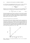
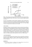
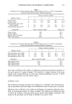
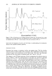
SALICYLIC ACID ENCAPSULATION 2 3 5 ß i ß i , i ß i i i 0 200 400 600 800 1000 1200 CONCENTRATION OF SA (lag/ml) Figure 4. Effect of the SA concentration in the donor compartment (0.2 M buffer solution) on the flux of SA across Silastic © membrane at pH 5 using a flow-through cell apparatus and a preserved saline (0.9% NaCI, 0.125% Chlorobutanol) receptor phase at 32øC under infinite dose conditions. EFFECT OF pH ON SALICYLIC ACID PERMEATION ACROSS SILASTIC © Under infinite dose conditions, an increase in pH dramatically decreases the permeation of SA through Silastic © membrane (Figure 5). The SA concentration used in this experiment was 0.550 mg/ml. The flux varies between 28.7 +-- 1.9 •g/cm2/h at pH 2 and 0.85 +- 0.02 •g/cm2/h at pH 4.5. This difference is flux can be related to the ionization of the SA at pH 4.5. The flux can be expressed as a sum of the contributions of the ionized and non-ionized forms: J = PsA ø CSA ø + Ps-Cs- (Eq. 9) lOO lO 1 .1 2 [D Flux Observed ß Flux Calculated ß [] ! ! ! i 3 4 5 6 pH of the Donor Solution Figure 5. Effect of the pH on the flux of SA across Silastic © membrane at 32øC from a 0.5-mg/ml SA solution in the donor compartment under infinite dose conditions. Calculated values are based on the only permeation of the nonionized form of salicylic acid with an estimated permeability coefficient of 49.9 x 10 - 3 cm/h. The calculation of the ionization of salicylic acid is based on Henderson-Hasselbalch equation assuming a pKa of 2.97, and the flux calculation is based on Fick's diffusion equation assuming sink conditions.

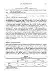
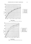
236 JOURNAL OF THE SOCIETY OF COSMETIC CHEMISTS PSA ø and Ps- are the permeability coefficients of the non-ionized and ionized forms, respectively, and Cs^ ø and C s- are the corresponding concentrations. Determination of the permeability coefficients was achieved by solving a system of equations involving the flux values obtained under different pH conditions. The cal- culated permeability coefficients are (49.9 --- 4.4) X 10 -3 cm/h for Ps^ ø and (0.06 --- 0.14) X 103 cm/h for Ps-- The latter was not significantly different from zero, which shows that only the unionized form was transported across the membrane. This agrees with a previous observation concerning the pH-partition hypothesis (20). Under infinite dose conditions, the diffusion profile of SA from a liposome preparation reached a steady state within an hour. The steady-state flux was obtained from the linear portion of the profile. An increase in phospholipid concentration (at an SA concentration of 0.5 mg/ml) at pH 4.5 decreased SA flux across the membrane (Figure 6). The flux dropped from 0.775 --- 0.035 •tg/cm2/h for a pH 4.5 buffer solution to 0. 125 --- 0.022 •tg/cm2/h for a 10% phospholipid preparation. Flux exhibited an inverse correlation with the entrapment of SA by the liposomes (Figure 6). These results suggest that an increase in phospholipid concentration reduced membrane flux by decreasing the SA activity in the donor. FINITE DOSE EXPERIMENTS Although transport from the pH 5 buffer through the membrane was initially very slow, a dramatic increase in penetration occurred as the preparation dried on the surface (Figure 7). Essentially all of the salicylic acid reached the receptor from the buffer during 120 100 80 60 40 20 Flux SA Bound i i i i i 0 2 4 6 8 10 60 50 40 30 20 10 0 12 Phospholipid Concentration (% w/w) Figure 6. Effect of phospholipid content of the donor phase on the entrapment of SA in phospholipid vesicles and on the flux of SA across Silastic © from a 0.5-mg/ml SA solution at pH 4.5 under infinite dose conditions.
Purchased for the exclusive use of nofirst nolast (unknown)
From: SCC Media Library & Resource Center (library.scconline.org)