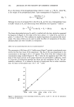
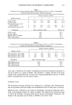
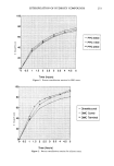
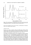
SALICYLIC ACID ENCAPSULATION 237 120 rO 200 HI BUFFER • I © 100 I•, BUFFER '• 100 I r• 200 I•1 5ø/o LIP. / • [,,• 4.::•i 100 HI 5% LIP. 1: 80 o 6o z ß • 40 o l: 20 d• .,...• 0 600 1200 1800 TIME IN MINUTES Figure 7. Effect of liposome entrapment of SA on its delivery across Silastic © under finite dose conditions. The initial preparation applied was a buffered solution of SA at pH 5. The donor compartment was open and subject to evaporation. The label "5% LIP." refers to a 5% w/w concentration of liposome material in the preparation. the time of the experiment (24 hours). However, application of SA in liposome prep- arations at the same pH served to decrease the penetration significantly so that only about 20% of the total amount of SA permeated during the same time period. Although an increase in the amount penetrated over time was noted, there was no sharp change in flux to correspond with that observed with the buffer. CONCLUSIONS Entrapment of salicylic acid could be described in terms of a partition-like equilibrium. The entrapment of SA in phospholipid vesicles is increased by lowering pH or increasing lipid concentration. Under infinite dose conditions in which excess material is applied, increasing entrapment results in a reduction in the steady-state flux of salicylic acid through an inert membrane. The effect of entrapment can be interpreted as a decrease in activity of SA in the preparation. There was also a reduction in penetration under finite dose conditions, which are more representative of typical skin applications, and in addition, the diffusion profile differed qualitatively from that of a buffer solution. ACKNOWLEDGMENTS The authors thank Richardson-Vicks, Division of Procter and Gamble, and American Lecithin for project support Dr. Deckner, Procter and Gamble, and Prof. Strauss, Rutgers University, for helpful discussions and Prof. Weyand, Rutgers University, for the use of equipment. REFERENCES (1) N. F. H. Ho, M. G. Ganesan, N. D. Weiner, and G. L. Flynn, Mechanism of topical delivery of liposomally entrappeal drugs, J. Controlled Release, 2, 61-65 (1985).

238 JOURNAL OF THE SOCIETY OF COSMETIC CHEMISTS (2) M. Mezei and V. Gulasekharam, Liposomes--A selective drug delivery system for the topical route of administration: Lotion dosage form, Li)• Sci, 26, 1473-1477 (1980). (3) W. Wohlrab and J. Lasch, Penetration kinetics of liposomal hydrocortisone in human skin, Derma- tologica, 174, 18-22 (1987). (4) J. Lasch and W. Wohlrab, Liposome bound cortisol: A new approach to cutaneous therapy, Biotaed. Biochim. Acta, 45, 1295-1299 (1986). (5) M. Mezei and V. Gulasekharam, Liposomes--A selective drug delivery system for topical route of administration: Gel dosage form, J. Pharm. Pharmacol., 34, 473-474 (1982). (6) K. Egbaria, C. Ramachandran and N. Weiner, Topical application of liposomally entrapped ciclo- sporin evaluated by in vitro diffusion studies with human skin, Skin Pharmacol., 4, 21-28 (1991). (7) N. Weiner, N. Williams, G. Birch, C. Ramachandran, C. Shipman, Jr, and G. Flynn, Topical delivery of liposomally encapsulated interferon evaluated in a cutaneous herpes guinea pig model, Antimicrob. Agents. Cheroother., 33, 1217-1222 (1989). (8) M. Jacobs, J.P. Martin, and C. Marriot, Effects ofphosphatidylcholine on the topical bioavailability of corticosteroids assessed by the human skin blanching assay, J, Pharm. Pharmacol., 40, 829-833 (1988). (9) A. Gesztes and M. Mezei, Topical anesthesia of the skin by liposome-encapsulated tetracaine, Anesth. Analg., 67, 107%1081 (1988). (10) W. Westerhof, Possibilities of liposomes as dynamic dosage form in dermatology, Medical Hypothesis, 16, 283-288 (1985). (11) M. Nakano and N. Patel, Release, uptake, and permeation behavior of salicylic acid in ointment bases, J. Pharm. Sci., 59, 985-988 (1970). (12) E. Gu6nin and J. Zatz, Interaction of skin with surfactant vesicle components, J. Soc. Cosmet. Chem. (in press). (13) A.D. Bangham, M. W. Standish, andJ. C. Watkins, Diffusion ofunivalent ions across the lamellae of swollen phospholipids, J. Mol. Biol., 13, 238-245 (1965). (14) R. L. Bronaugh and R. F. Stewart, Methods for in vitro percutaneous absorption studies. IV. The flow-through diffusion cell, J. Pharm. Sci., 74, 64-67 (1985). (15) Merck Index, Eleventh Edition (Merck & Co. Inc., 1989). (16) A. Martin, J. Swatbrick, A. Cammarata, and A. Chun, Physical Pharmacy, Third Edition (Lea & Febiger, Philadelphia, 1983). (17) M. Grit, J. H. De Smidt, A. Struijke, and D. J. A. Crommelin, Hydrolysis ofphosphatidylcholine in aqueous liposome dispersions, Int. J. Pharm., 50, 1-6 (1989). (18) W. I. Higuchi and T. Higuchi, Theoretical analysis of diffusional movement through heterogeneous barriers, J. Am. Pharm. Assoc. Sci., 49, 598-606 (1960). (19) G. L. Flynn and T. J. Roseman, Membrane diffusion. II. Influence of physical adsorption on mo- lecular flux through heterogeneous dimethylpolysiloxane barriers, J. Pharm. Sci., 60, 1788-1796 (1971). (20) E. R. Garrett and P. B. Pramod, Evaluation, control, and prediction of drug diffusion through polymeric membranes: A test of the pH-partition hypothesis, J Pharm. Sci., 57, 949-959 (1968).
Purchased for the exclusive use of nofirst nolast (unknown)
From: SCC Media Library & Resource Center (library.scconline.org)