SURFACTANT VESICLES 79 added, and the lipids were hydrated at a temperature above their transition temperature. A 5% phospholipid concentration (weight/weight) was achieved after buffer addition. The manufacture was completed by a brief sonication to provide homogeneity of the preparation. Nonionic surfactant vesicles (NSV) were prepared using the same buffers, employing high shear mixing as per U.S. patent 4,855,090. Briefly, a mixture of nonionic sur- factant, cholesterol, and charge additive was heated at a temperature above the transition temperature of the highest melting component in the mixture. The buffer solutions were heated separately to the same temperature as the lipophilic phase. Aqueous and li- pophilic phases (80:20 w/w, respectively) were rapidly introduced into two different syringes connected by a three-way Luer-Lock © valve. The content of the syringe was then forced into the second syringe through an orifice about 1 mm in diameter. The resulting mixture was driven back and forth between the two syringes until the prep- aration was brought back to room temperature. The presence of vesicles in the prepa- ration was observed using freeze-fracture 'scanning electron microscopy. PROTOCOL OF INVESTIGATION To investigate the effect of the preparation, the hairless mouse skin, free of holes or defects (visual inspection), was mounted on the diffusion cell of the flow-through diffusion apparatus (Vanguard International, Neptune, NJ) (12). The diffusion cells, with a cross-sectional area of 0.64 cm 2, were maintained at 32øC by a water-circulating system. 200 p,1 of the vesicle preparations made of phospholipids or nonionic surfac- tants, or 200 p,1 of a control, was applied to the skin in the donor compartment of the vertical cell and allowed to remain in place for a period of 12 hours. The receptor side of the cell contained preserved saline solution (NaC1 0.9%, chlorobutanol 0. 125%), flowing through at a rate of 5 ml/h. Occlusive conditions, when desired, were achieved by covering the cell with a layer of Parafilm ©. After the period of application, the surface of the skin was washed with an alcohol/water (1:2) mixture until it appeared cleaned of the preparation. Then 200 p,1 of water spiked with tritiated water was applied, and the permeation of the radiolabeled water was monitored under occlusive conditions, with fraction collection, for a period of three hours. The cell was then dismantled, and the donor phase, washing fraction, and collected fraction were counted. The water perme- ation rate (WPR) was calculated at steady state and reported in p,1/h. Each step of this experiment was conducted using four replicates. The results are reported with the standard deviation (SD). SCINTILLATION COUNTING Liquid scintillation cocktail (Scintiverse TM BOA, Fisher Scientific, Fair Lawn, NJ) was added, and the radioactivity in the vials was counted for a ten-minute period to obtain a precise estimate of the [3HI water permeated. The scintillation counter was a Beckman LS 5000 TD (Beckman Instruments, Fullerton, CA). Previously a quench curve had been established using acetone as a quenching agent, and the integrated software con- verted the count per minute (CPM) to disintegrations per minute (DPM), using the H number.

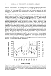
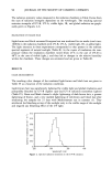
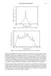
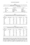
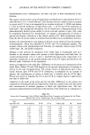
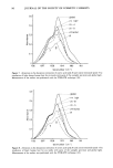
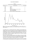
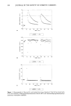
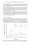
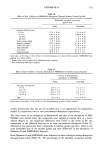
80 JOURNAL OF THE SOCIETY OF COSMETIC CHEMISTS RESULTS AND DISCUSSION EFFECT OF pH ON SKIN DAMAGE The permeation of tritiated water reached a steady state after approximately 20 minutes and showed no depletion effect during the experiment, except in the case of sodium lauryl sulfate pretreatment. All the water permeation rates (WPR) were compared to the original permeation rate through freshly excised mouse skin with no treatment (WPR: 1.34 - 0.25 [•l/h). Permeation profiles (Figure 1) showed a small damaging effect of the pH 2 buffer under closed and open conditions (WPR: 5.09 - 0.68 and 3.04 - 0.25 [•l/h, respectively), but very little effect of the pH 5 buffer in both application condi- tions (WPR: 1.91 - 0.23 [•l/h under occluded conditions and 1.11 - 0.15 under open conditions of treatment). All buffer treatments, except pH 5 under open conditions, resulted in WPR values statistically significantly different from no treatment (two tails t-test with 95% confidence interval). The damaging effect of the pH 2 buffer is con- sistent with the corrosive and denaturing properties of strong acids. Values obtained with buffer solutions were used as references for the studies with surface active com- pounds. SODIUM LAURYL SULFATE Sodium lauryl sulfate as a I% solution was introduced as a positive control. No attempt was made to control the pH of this solution. The significant water permeation, which resulted from the application of this positive control (Figures 2-5), was a sign of skin damage, which is consistent with a previous report (5). The skin damage was more 20 10 ,& CLOSED pH 2 A OPEN pH 5 {• CLOSED O OPEN • FRESH,SKIN ..... ß. •. •,•,• •1 •-'.,•..-" I ' I ' I ' I 0 I 2 3 4 TIME IN HOURS Figure 1. Effect of treatment by pH 2 and pH 5 buffers on the water permeation across hairless mouse skin as compared to a freshly excised and nontreated skin sample (open vs occluded application). The error bars represent the standard deviation.
Purchased for the exclusive use of nofirst nolast (unknown)
From: SCC Media Library & Resource Center (library.scconline.org)