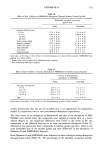
PADIMATE-O 119 Viability of skin was maintained in the diffusion cells for 24 h by using a HEPES- buffered Hank's balanced salt solution (HHBSS) with the addition of gentamycin sulfate and bovine serum albumin (9). In some experiments, sodium fluoride was added to the above receptor fluid to render the skin nonviable (10). All receptor fluids were sterilized before use by passage through a 0.2-lxm filter (Nalgene Company, Rochester, NY). A simple oil-in-water emulsion lotion was prepared for use as one of the vehicles in this study. The stearic acid (3%) and water (91%) emulsion contained 4% Padimate-O to simulate a sunscreen product and 1% propylene glycol as a humectant. Radiolabeled Padimate-O in both an ethanol and a lotion vehicle was applied to the skin (15 }xl/cm 2) at a chemical dose of 6.7 }xg/cm 2. Radiolabeled NMPABAO in the lotion was applied to the skin (15 [•l/cm 2) at a chemical dose of 7.0 }xg/cm 2. The skin was not occluded therefore, the vehicles were allowed to evaporate as they would during con- ditions of normal use. All skin surfaces were washed at 24 h with a 1% solution of soap (dishwashing detergent) and water to remove unabsorbed material. Test compound not removed from the skin by washing was considered to be absorbed and was added to the receptor fluid levels to determine total percutaneous absorption. Localization of compounds in skin was measured by stripping each skin section ten times with cellophane tape to remove the stratum corneum. Skin sections were then homog- enized in HHBSS solution by using a Polytron tissue homogenizer (Brinkmann Instru- ments, Westbury, NY). Small aliquots (0.2 ml) of the receptor fluid (collected at 6-h intervals), tape strips, and skin homogenates were analyzed for radioactivity by using a Beckman LS9000 scintillation counter (Beckman Instruments, Irving, CA). Parent compound and metabolites were extracted from the remaining receptor fluid and skin homogenates by using Sep-Pak C •g cartridges (Waters Associates, Milford, MA). All receptor fluids and skin homogenates were adjusted to pH 3.2-3.5 and filtered through the cartridges. Compounds bound to the Sep-Pak cartridges were eluted in 4 ml of acetone. The eluates containing parent compound and metabolites were applied to silica gel thin-layer chromatography (TLC) plates along with nonradiolabeled standards of Padi- mate-O (ICI Americas Inc., Wilmington, DE), dimethyl aminobenzoic acid (DMABA) (Aldrich Chemical Corp., Milwaukee, WI), NMPABAO, and p-(N-methyl-N-nitros- amino) benzoic acid (NMPABA) (Cosmetics Technology Branch, FDA, Washington, DC). TLC plates were developed with hexane:ethyl acetate (135'15) as the solvent system. Radioactivity on TLC plates was quantified by a Bioscan TLC plate scanner (Bioscan Inc., Washington, DC). Radioactivity in each spot was expressed as the percent of total radioactivity applied to the plate. Re-values corresponding to the radioactive peaks were compared with those of standard compounds. Initial photodecomposition studies were performed by spreading 300 mg of a commer- cial sunscreen product containing 800 ppb NMPABAO to a thickness of approximately 20 }xm over the bottom of a 20 x 150-ram petri dish. The petri dish containing the test portion was exposed to a Mutzhas UV light source with a Supuvasun 3000 filter (Mutzhas, Munich, Germany) at a distance of 207 cm from the light source. At this distance, UVA and UVB output readings averaged 6.7 x 10 -2 and 0.40 x 10 -2 mW/cm 2, respectively. Test portions were exposed for 0.0, 1.0, 1.5, and 2.0 rain, with two repetitions for each time period. For the 2.0-rain time interval, the UVB radiation dose was 0.48 mJ/cm 2. The human minimal erythema dose (MED) required to produce

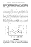
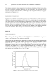
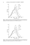
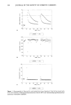
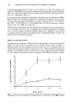
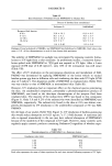
120 JOURNAL OF THE SOCIETY OF COSMETIC CHEMISTS a minimal perceptible redness on skin is 20-50 mJ/cm 2 (11). After UV exposure, the amount of NMPABAO remaining was determined by high-pressure liquid chromatog- raphy with nitrosamine-specific thermal energy analyzer detection (Thermedics Detec- tion, Inc., Chelmsford, MA)(12). In some percutaneous absorption experiments, after application of radiolabeled NMPA- BAO, the skin was immediately exposed to UV light from the Mutzhas solar simulator for 3 min before initiation of the diffusion cell study in darkness (UVA = 0.67 mW/cm 2 and UVB = 0.028 mW/cm2). At this radiation dose, the skin was exposed to approx- imately 25% of the MED for human skin. Absorption and metabolism data were analyzed for significance by using the Student's t test to compare means for two test samples (Instat, Graphpad Software, San Diego, CA). A one-way analysis of variance (ANOVA) followed by a Bonferroni test was used to compare means for multiple test samples (Instat, Graphpad Software). P-values less than 0.05 were considered significant. RESULTS AND DISCUSSION The percutaneous absorption of Padimate-O was measured from a lotion and an ethanol vehicle the time course of absorption into the receptor fluid is shown in Figure 1. Absorption from the lotion appeared to reach a steady state at 6 h absorption from the ethanol vehicle appeared to reach a steady state at 12 h. Values obtained for absorption from the lotion through viable and nonviable skin were not significantly different (Table I). This comparison demonstrates that the integrity of the diffusional barrier to absorp- m 5 m 4 o o 6 TIME (hours) Figure 1. Time course of percutaneous absorption of Padimate-O in hairless guinea pig skin. Padimate-O was applied to skin in 15 Ixl vehicle/cm 2 at a chemical dose of 6.7 Ixg/cm 2. ß = Lotion ß = Ethanol.
Purchased for the exclusive use of nofirst nolast (unknown)
From: SCC Media Library & Resource Center (library.scconline.org)