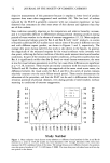
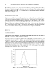
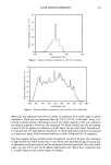
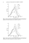
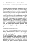
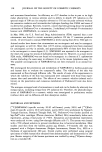
SURFACTANT VESICLES 83 lOO 80 60 40 20 TREATMENTS [] SLS [] LIPOSOME [] NSV BUFFER , Figure 4. Water permeation rate across hairless mouse skin after different treatments at pH 5 under open conditions. (Sodium lauryl sulfate solution was prepared in deionized water and was not pH-controlled.) The error bars represent the standard deviation. 80 70 60 50 40 30 20 10 0. TREATMENTS [] SLS [] LIPOSOME [] NEUTRAL NSV 1• BUFFER ß Figure 5. Water permeation rate across hairless mouse skin after different treatments at pH 5 under closed conditions. (Sodium lauryl sulfate solution was prepared in deionized water and was not pH-controlled.) The error bars represent the standard deviation. EFFECT OF APPLICATION CONDITIONS The type of application, that is whether the skin surface is occluded or not, affects water transport rate through the skin. As shown above, occlusion sometimes results in an increase and sometimes a decrease in WPR, depending on the nature of the system applied. The increase of surfactant concentration during the pretreatment in open conditions did not increase the WPR dramatically, as is the case with a skin-damaging agent such as SLS.

84 JOURNAL OF THE SOCIETY OF COSMETIC CHEMISTS CONCLUSION Using the water permeation rate as an indicator, pretreatment with nonionic surfactant vesicle preparations at either pH 2 or 5 does not damage hairless mouse skin. The same is true for phospholipid-based vesicles at pH 5, but not at pH 2. REFERENCES (1) N. Weiner, N. Williams, G. Birch, C. Ramachandran, C. Shipman, and G. Flynn, Topical delivery of liposomally encapsulated interferon evaluated in a cutaneous herpes guinea pig model, Antimicrob. Agents Cheroother., 33, 1217-1221 (1989). (2) M. Jacobs, J.P. Martin, and C. Marriot, Effects ofphosphatidylcholine on the topical bioavailability ofcorticosteroids assessed by human skin blanching assay,J. Pharm. Pharmacol., 40, 829-833 (1988). (3) K. Egbaria, C. Ramachandran, D. Kittayanond, and N. Weiner, Topical delivery of liposomally encapsulated interferon evaluated by in vitro diffusion studies, Antimicrob. Agents Chemother., 34, 107-110 (1990). (4) W. Westerhof, Possibilities of liposomes as dynamic dosage form in dermatology, Medical Hypothesis, 16, 283-288 (1985). (5) M. Loden, The simultaneous penetration of water and sodium lauryl sulfate through isolated human skin. J. Soc. Cosmet. Chem., 41, 227-233 (1990). (6) J. A. Faucher and E. D. Goddard, Interaction ofkeratineous substrates with sodium lauryl sulfate: II. Permeation through stratum corneum, J. Soc. Cosmet. Chem., 29, 339-352 (1978). (7) R. Bronaugh, R. F. Stewart, and M. Simon, Methods for in vitro percutaneous absorption studies. VII: Use of excised human skin. J. Pharm. Sci., 75, 1094-1100 (1986). (8) H. E. J. Hofland, J. A. Bouwstra, J. C. Verhoef, G. Buckton, B. Z. Chowdry, M. Ponec, and H. E. Junginger, Safety aspects of non-ionic surfactant vesicles: A toxicity study related to the physico-chemical characteristics of non-ionic surfactants, J, Pharm. Pharmacol., 44, 287-294 (1992). (9) H. E. Hofland, Vesicles as transdermal drug delivery systems, Ph.D. Thesis, Leiden University, The Netherlands (1992). (10) K. A. Walters, M. Walker, and O. Olejnik, Non-ionic surfactant effects on hairless mouse skin permeability characteristics, J. Pharm. Pharmacol., 40, 525-529 (1988). (11) A.D. Bangham, M. M. Standish, andJ. C. Watkins, Diffusion ofunivalent ions across the lamellae of swollen phospholipids, J. Mol. Bid., 13, 238-252 (1965). (12) R. L. Bronaugh and R. F. Stewart, Methods for in vitro percutaneous absorption studies. IV: The flow-through diffusion cell, J. Pharm. Sci., 74, 64-67 (1985). (13) M. Grit, J. H. de Smidt, A. Struijke, and D. J. A. Crommelin, Hydrolysis ofphosphatidylcholine in aqueous liposome dispersions, Int. J. Pharmaceut., 50, 1-6 (1989).
Purchased for the exclusive use of nofirst nolast (unknown)
From: SCC Media Library & Resource Center (library.scconline.org)