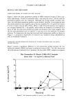
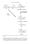
86 JOURNAL OF THE SOCIETY OF COSMETIC CHEMISTS studied the dermal penetration and systemic distribution of [•4C]-labeled vitamin E and reported that vitamin E penetrated the skin very well and that a high accumulation of radioactivity in the skin was observed (6). Until now, skin penetration studies of vitamin E have been performed with radiolabeled vitamin E, and the total radioactivity through the skin has been assayed. Since a major route of skin penetration is through lipid-rich intercellular routes, drugs that have good lipid solubility and favorable oc- tanol/water partition coefficients transfer into the skin readily (7). Based on the above studies, it has been generally concluded that vitamin E penetrates the skin very well. However, we observed that very lipophilic vitamin E (P. C. octanol/water = 476) did not appear in the receptor solution after quite a long lag time and showed a very small permeation rate in HPLC analysis. The significant difference in the penetration profile was observed between HPLC assay and radiotracer analysis. The radiolabeled vitamin E promptly penetrated the skin, while the nonlabeled compound appeared after a remark- able long time lag (about 24 hours). Although there is some evidence for the existence of vitamin E metabolites such as tocopherol quinone, tocopherol hydroquinone, dimers, and trimers in other tissues (8-10), no metabolism study for vitamin E in the skin has been reported. To investigate the possible explanation for these observation, the metabolism of vitamin E in hairless mouse skin has been investigated. MATERIALS AND METHODS Vitamin E (o•-tocopherol), o•-tocopherol quinone, and radiolabeled vitamin E (•[H] 2- dl-tx-tocopherol, 2.0 mCi/mM) were kindly provided by Hoffmann-LaRoche (Nutley, NJ). The purity of radiolabeled compounds was tested by TLC or HPLC in line with a liquid scintillation counter (Rack Beta 1214-001, LKB Instruments Inc., Gaithersburg, MD). Tween-80 (ICI Americas, Inc., Wilmington, DE) and silicone fluid (Dow Corn- ing 360, 20 cp) were used as obtained. All other chemicals were reagent grade. The solvent used in the HPLC assay was HPLC grade. Water was purified by a Nanopure water purification system (Sybron/Barnstead, Boston, MA). Bioflour (E.I. Dupont NEN Research, MA) was used as a liquid scintillation cocktail. ANALYTICAL METHODS HPLC analysis. When the mobile phase of methanol:chloroform (90:10) was used for vitamin E analysis, the vitamin E peak was very sharp at a retention time of about two minutes. To separate vitamin E and its metabolites, we increased the water fraction of the mobile phase to prolong the retention time, resulting in better separation of vitamin E from its metabolites. Methanol:water (95:5) was selected as a mobile phase. The retention time of vitamin E under this condition was about 12 minutes. The assay conditions are summarized in Table I. Radioactivity counting. A liquid scintillation counter (LSC) was used to quantify the concentration of radiolabeled vitamin E. For the assay, the samples (30-80 Ixl) were withdrawn and mixed well with Bioflour scintillation cocktail (5-10 ml). The amount of radioactivity (disintegration per minute, dpm) was determined, and the correction for quenching was made automatically by comparison against a standard quench curve.

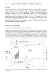
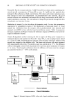
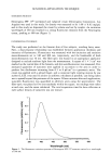
VITAMIN E METABOLISM 87 Table I Assay Condition of Vitamin E with HPLC in Line With a Liquid Scintillation Counter Flow rate Mobile phase (ml/min) Detection wavelength Column type MeOH:water = 95:5 1.0 280 nm •-bondapak C18(RP), 4 X 30 mm HPLC system: pump Waters 510 injector WatersWISP 710B detector Kratos Spectroflow 773 integrator Waters 730 data module. LSC system: B-Ram Micropump (CA) elution buffer Bioflour (1 ml/min). Time was calculated from the corrected values of dpm and the specific activity (dpm/mg) of radiolabeled vitamin E in the donor solution at time zero and the concentration of vitamin E in the receptor solution at each sampling. Permeation study. A freshly excised full thickness of abdominal skin of a female hairless mouse (5-7 weeks old, Jackson Lab. HRS/J Strain) was mounted between the half cells of the in vitro skin permeation system, as shown in Figure 1. Vitamin E (13 mg/ml) or [3H]2-dl-tx-tocopherol with vitamin E in silicone fluid was employed as a donor solu- tion. An aqueous solution of Tween-80 (5 mM) was used as a receptor solution. The solubility of vitamin E in silicone fluid was 39 mg/ml. To increase the solubility of vitamin E in receptor solution, maintaining a sink condition during the permeation experiment, Tween-80 was used. Tween-80 can increase the solubility of the lipophilic compound by incorporating vitamin E into a micelle. Imai et al. (11) reported that the solubility of vitamin E in water increased linearly with increasing concentration of ......•j.•...- Glass stopper Sampling port .•,• •. • • to other cells 37øC water in • '-"•:•i- (in series) skin Receptor compartment (3.5 ml ) Donor compartment • (:•5 ml) Water- (37 ø ) Star- head magnet (dia.,8.5 mm height,6 ram) Stirring platform (dia., 10 ram) (height, 4mm) Revolving magnet (6oo rpm) •ynchronous motors -- • Connecting • on-off button tubing SKIN PERMEATION SYSTEM by VALIA & CHIEN Figure 1. Schematic diagram of a Valia-Chien diffusion cell for skin permeation study.
Purchased for the exclusive use of nofirst nolast (unknown)
From: SCC Media Library & Resource Center (library.scconline.org)