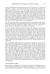
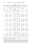
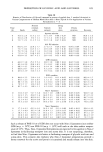
DEPOSITION OF GLYCOLIC ACID AND GLYCEROL 107 Table V Kinetics of Distribution of Glycolic Acid (expressed as percent of applied dose + standard deviation) in Various Strata of Hairless Mouse Skin After 1-Hour Topical In Vivo Application of Nonionic Liposomal Formulations Containing 2.5% Glycerol (n = 3) Stratum Living Time corneum Stratum skin Urinary (h) Swabs surface corneum strata excretion Recovery Non-1 liposomes with 2.5% glycerol 0 46.4 + 5.6 33.9 + 4.1 5.8 + 0.7 2.02 + 0.30 0.05 + 0.02 88.1 + 0.8 4 29.3 + 1.2 22.3 + 0.7 13.8 + 0.5 1.40 + 0.19 1.20 + 0.22 68.0 + 0.5 8 36.7 + 2.8 18.3 + 0.4 10.3 + 1.8 0.72 + 0.13 1.00 + 0.08 67.0 + 2.3 Non-2 liposomes with 2.5% glycerol 0 20.5 + 2.7 61.7 + 0.3 8.9 + 3.0 1.04 + 0.06 0.08 + 0.03 90.0 + 1.2 4 22.5 + 2.8 34.2 + 0.7 14.9 + 1.9 1.48 + 0.16 0.36 + 0.02 73.4 + 2.8 8 19.4 + 5.3 23.2 + 6.2 14.9 + 3.8 1.24 + 0.04 0.86 + 0.42 59.6 + 3.3 stratum corneum, in comparison with conventional vehicles. Further, the greater amounts of glycolic acid found in the living skin strata from these nonionic liposomal formulations indicate their utility in transporting actives to living tissue for therapeutic effects. The results also strongly suggest that of the two nonionic liposomes tested, Non-2 liposomes may allow increased retention of glycolic acid at the site of application without enhancing percutaneous absorption. REFERENCES (1) L. Overgaard-Olsen and G. B. Jemec, The influence of water, glycerin, paraffin oil and ethanol on skin mechanics, Acta. Derre. Venereal (Stockh), 73, 404-406 (1993). (2) E. J. Van Scott and R. J. Yu, nyperkeratinization, corneocyte cohesion and alpha-hydroxy acids, J. Am. Acad. DermatoL, 11, 867-879 (1984). (3) E. J. Van Scott and R. J. Yu, Control of keratinization with alpha-hydroxy acids and related com- pounds. I. Topical treatment of ichthyotic disorders, Arch. Dermatol., 110, 586-590 (1974). (4) M. Goldstein and R. Brucks, Evaluation of glycolic acid permeation through skin, Pharm. Res., 11, S-180 (1994). (5) C. Ackermann and G. L. Flynn, Ether-water partitioning and permeability through nude mouse skin in vitro. I. Urea, thiourea, glycerol and glucose, lnt. J. Pharm., 36, 61-66 (1987). (6) A. Rougier, D. Dupuis, C. Lotte, R. Roguet, and H. Schaefer, In vivo correlation between stratum corneum reservoir function and percutaneous absorption, J. Invest. Dermatol., 81, 275-278 (1983). (7) L. Stryer, Biochemistry, 3rd ed. (W.H. Freeman, New York, 1988), p. 392. (8) S. M. Dowton, Z. Hu, C. Ramachandran, D. F. H. Wallach, and N. Weiner, Influence of liposomal composition on topical delivery of encapsulated cyclosporine-A. I. An in-vitro study using hairless mouse skin, STP Pharma Sci., 3, 404-407 (1993). (9) S. M. Niemiec, Z. Hu, C. Ramachandran, D. F. H. Wallach, and N. Weiner, The effect of dosing volume on the disposition of cyclosporine-A in hairless mouse skin after topical application of a nonionic liposomal formulation: An in vitro diffusion study, STP Pharma Sci., 4, 145-149 (1994). (10) P. P. Sarpotdar and J.L. Zatz, Evaluation of penetration enhancement of lidocaine by nonionic surfactants through hairless mouse skin in vitro, J. Pharm. Sci., 75, 176-181 (1986). (11) K. A. Walters, M. Walker, and O. Olejnik, Non-ionic surfactant effects on hairless mouse skin permeability characteristics, J. Pharm. Pharmacol., 40, 525-529 (1987).

j. Soc. Cosmet. Chem., 47, 109-115 (March/April 1996) Quantitative assessment of sunscreen application technique by in vivo fluorescence spectroscopy L. E. RHODES and B. L. DIFFEY, Regional Medical Physics Department, Dryburn Hospital, Durham DH1 5TW, United Kingdom. Received September 1995. Synopsis For the first time, a method is described for measurement of surface density of sunscreen in vivo. Here, the method is used to measure the uniformity of sunscreen application. The intrinsic fluorescence of a sunscreen (Neutrogena SPF 15 ©) was quantified by fluorescence spectroscopy, and a dose-response relationship was established with sunscreen density on the skin. Sunscreen was then applied in a crude fashion to one forearm and carefully to the other forearm in five subjects. Fluorescence measurements were taken from 16 sites on each forearm and converted to an equivalent thickness of sunscreen using the dose-response relationship. Whereas the median thicknesses for crude and careful application were approximately the same, the range of thickness was higher after crude application (p 0.007). Hence fluorescence spectroscopy can quantify the adequacy of sunscreen application. This simple and rapid noninvasive in vivo technique for measuring sunscreen thickness could potentially provide a surrogate method for SPF determination in the clinical testing of new products. INTRODUCTION New sunscreens are developed with the aim of protecting the skin from sunburn and chronic photodamage. The protection offered by a sunscreen is assessed by the sun protection factor (SPF) following phototesting in vivo. Sunscreen efficacy is obviously dependent on appropriate application. Current laboratory testing of sunscreen efficacy is based on an even application thickness of 2mg/cm 2. This surface density of sunscreen is recommended by the U.S. Food and Drug Administration (1), the Standards Association of Australia (2), and the International Commission on Illumination (3), while the German Standards Organisation Deutsches Institut ffir Normung recommends the slightly lower surface density of 1.5 mg/cm 2 (4). However, crude assessments of application technique in a more natural setting suggest consumers apply a much lower average sunscreen thickness of 0.5-1.3 mg/cm 2. A number of investigators have determined the average layer thickness of sunscreen applied L. E. Rhodes' present address is: Dermatology Unit, University of Liverpool, P.O. Box 147, Liverpool L69 3BX, U.K. 109
Purchased for the exclusive use of nofirst nolast (unknown)
From: SCC Media Library & Resource Center (library.scconline.org)