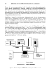
110 JOURNAL OF THE SOCIETY OF COSMETIC CHEMISTS by subjects by weighing the sunscreen containers before and after application, and then dividing the amount applied by the area treated. Using this method, Stenberg and Lark/5 (5) found that the average thickness of applied sunscreen was around l mg/cm 2 and that this varied little with region of the body or product. Minimal erythema dose (MED) testing then demonstrated that the SPF on areas of self-application of the cream was only 50% of that on areas given the generally recommended thickness of 2 mg/cm 2. Gottlieb et al. (6) also found the average application thickness at l. 3 mg/cm 2 to be nearer 1 than 2 mg/cm 2. Bech-Thomsen & Wulf (7) asked subjects to apply their own sunscreen to their whole body, in a beach setting. In this study, the amount of sunscreen applied was on average only 0.5 mg/cm 2. Some authors have studied application technique following the addition of fluorescent substances to sunscreens. The presence or absence of the sunscreen has then been confirmed by detection of fluorescent emission following irradiation with Wood's light, with peak excitation wavelength around 365 nm. Stenberg and Lark/5 (5) added 10% dihydroxyacetone as a fluorescent agent, while Loesch and Kaplan (8) employed doxy- cycline. In the latter study, subjects were asked to apply the sunscreen to their faces, and absence of fluorescence showed that certain sites tended to be completely missed. In the above studies, only the gross average density of sunscreen applied could be calculated, and the fluorescent emission of sunscreens was used in a purely qualitative manner. Sauermann and Hoppe (9) attempted to use fluorescence spectrometry for indirect assessment of sunscreens, observing attenuation of fluorescence of a dye, dansyl chloride, by the subsequent application of sunscreen. Fluorescence spectroscopy enables the use of a wide range of excitation and emission wavelengths, with quantification of the intensity of fluorescent radiation (10). These instruments are designed primarily for in vitro use, but by coupling suitable fiber optics, the excitation radiation may be conducted to remote sites and the emission radiation conducted back to the spectrometer (l 1). Hence the technique, which can employ either a laser or incoherent light source, has been used in the identification of atheromatous plaque (12) and abnormal gastric mucosa (13), and more recently, in the study of skin fluorophores. Differences have been described in the fluorescence patterns of photoaged and chronologically aged skin (14) and at the site of melanomas (15). We have now extended previous studies by examining the variation of sunscreen appli- cation technique in a quantitative manner, exploiting the intrinsic fluorescence of a sunscreen. MATERIALS AND METHODS MEASUREMENT OF FLUORESCENCE A fluorescence spectrometer (Fluoromax, Spex Industries Inc.) was adapted for use in this study. The radiation from a xenon arc lamp was focused into the excitation mono- chromator. The spectrometer was coupled to a bifurcated fiber-optic cable, enabling conduction of excitation radiation to the skin and collection of fluorescent radiation, which was directed into the entrance slit of the emission monochromator. The signals were processed and the data displayed using the computer software.

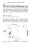
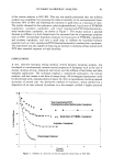
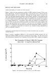
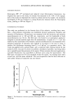
SUNCREEN APPLICATION TECHNIQUE 111 SUNSCREEN PRODUCT Neutrogena SPF 15 © waterproof and rubproof cream (Neutrogena Corporation, Los Angeles) was used in this study. Its density was measured to be 1.08 __ 0.02 mg/txl, and in this study we dispensed the cream by volume and not by weight. An excitation wavelength of 340 nm resulted in a strong fluorescent emission from the Neutrogena cream, peaking at 400 nm (Figure 1). EXPERIMENTAL TECHNIQUE The study was performed on the forearm skin of five subjects, avoiding hairy areas. First, a dose-response relationship was established between application thickness and intensity of fluorescence. Fluorescence was measured with the excitation and emission monochromators set to 340 and 400 nm, respectively, and the bandwidth of both monochromators was 10 nm. The fiber-optic was housed in an appropriate applicator designed to exclude ambient light from the measurement. A square of 5 x 5 cm 2 was marked on the ventral skin of the forearm, and skin auto fluorescence was measured. Five measured quantities of sunscreen were applied in succession to the area in order to produce five thicknesses increasing from 0.5 to 8 txl/cm 2 in a geometric series. The cream was applied with a gloved finger, and a consistent light rotating motion by one author (L. E. R.) was used to achieve as uniform a thickness as possible, care being taken not to spread beyond the perimeter of the square. Fifteen minutes after each application, sunscreen fluorescence was quantified in arbitrary units using the spectrometer. The fiber-optic was placed at five sites randomly within the square, ten readings were taken at each site, and the mean calculated. The total acquisition time for data collection at each surface density of sunscreen was one minute. 0.8 0.6 0.4 0.2 * I ' I I I I •50 400 450 500 550 600 Wavelength (nm) Figure 1. The fluorescence emission spectrum of Neutrogena SPF 15 © sunscreen excited at a wavelength of 340 nm.
Purchased for the exclusive use of nofirst nolast (unknown)
From: SCC Media Library & Resource Center (library.scconline.org)