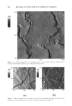
EFFECT OF ANTI-KERATIN ANTIBODY ON HAIR 211 of the wells (MultiScreen plate, Millipore Ltd.). Five-micrometer-thick sections were sliced off from human hair with a cryostat. Sections of fifteen hundreds were put in each of the wells. The plate was then blocked for one hour at room temperature with 3% bovine serum albumin (Seikagaku Kogyo Co.) in PBS. It was then washed twice with 0.02% Tween©-20 in saline. Each dilution (100 lal) of the anti-keratin antibody or non-specific antibody was applied to each well and incubated for one hour at room temperature. After the wells were washed five times, rabbit anti-antibody peroxydase conjugate (Kappel) that was diluted 1:500 was added (100 lal) to each well and incubated for one hour. Wells were washed again five times and 0.4 mg/ml 0-phenylenediamine in 0.1 M Na2HPO4, 0.05 M citric acid, and 0.006% H202 was added (tOO lal/well). After 30 min, 3N H2SO 4 was added to stop the coloring. The amount of color was read at 492 nm with a microplate reader (MTP-32, Colona Electric Co., Ltd.). In the binding assay of the antibody to hair fiber, a lock of tOO hair strands was immersed in PBS containing 0.3% antibody for one hour at 30øC. The strands were thoroughly rinsed with t000 ml of 0.05% Tween©-20 in PBS (PBS-T) and then fixed with 0.25% glutaraldehyde, rinsed with PBS-T, and sliced into sections. To detect the amount of antibody binding to hair fibers, application of ELISA to the hair sections was performed according to the above-mentioned method. TENSILE PROPERTIES In order to reduce the deviation attributed to the inequality of the diameter of each hair, two adjoining sections (7 cm) of the same hair were used as a pair. One of the two sections was immersed in PBS containing 0.3% antibody for one hour at 30øC and another in PBS. These fibers were then rinsed with tap water for ten minutes. Stress- strain curves for individual hair fibers were obtained at 22øC and 65% relative humidity using the tensile tester. The hair fiber (5 cm) was stretched at a rate of 5 cm/min. The elastic modulus (Es), (the ratio of stress to strain) in the Hookean region, was calculated as• Es = AF * L/A * AL (N/M 2) where AF = the change in force induced by a change in length, L = fiber length in meters, AL -- fiber extension in meters, and A = fiber cross-sectional area in meters squared, which was determined by the weight per length, supposing the density is t g/cm 3. We evaluated the stress to breaking point (the tensile strength) and the percentage extension to breaking point as other parameters of load elongation curves. For statistical analysis of those parameters, we used paired t-tests. GENERATION OF FRACTURE BY BRUSHING Two grams of hair strands (15 cm in length) were immersed in PBS containing 0.3% antibody, its fragments, or non-specific antibody for one hour at 30øC. As a control, a hair lock was immersed in PBS. The hair was rinsed with tap water for ten minutes after a hundred brush strokes. Before brushing, the hair lock was immersed in water for one minute and was partially dried by using a drier for one minute in order to accelerate the damage to hair by brushing. The hair was brushed manually using the blind study method. The number of fractures generated in hair locks was counted.

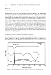
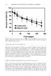
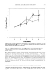
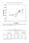
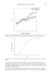
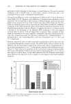
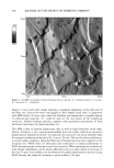
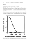
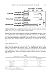
212 JOURNAL OF THE SOCIETY OF COSMETIC CHEMISTS RESULTS BINDING ABILITY OF THE ANTI-KERATIN ANTIBODY TO HAIR FIBER The binding ability of the anti-keratin antibody to hair sections is shown in Figure 1. The absorbance indicates that binding of antibodies to hair sections was increased in proportion to the concentration of the anti-keratin antibody, but not of non-specific antibody. This result indicates that the anti-keratin antibody does possess the ability to bind to hair. The abi'lity of the antibody to bind to the damaged hair fiber is shown in Figure 2. No difference was observed in the amount of non-specific antibody binding to virgin hair or to damaged hair. On the other hand, the binding of the anti-keratin antibody was 1.5 o.5 1000 100 10 ! Concentration of Antibody (ng/ml) Figure 1. The binding ability of antibody to hair sections. The binding of antibody to hair sections was detected by using an indirect ELISA method. Hair sections were incubated with the antibody (C)) and with non-specific antibody (O).
Purchased for the exclusive use of nofirst nolast (unknown)
From: SCC Media Library & Resource Center (library.scconline.org)