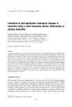
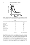
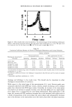
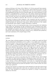
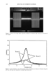
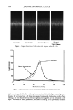
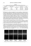
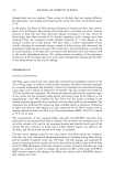
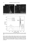
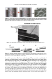
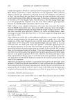
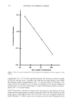
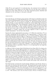
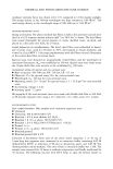
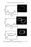
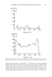
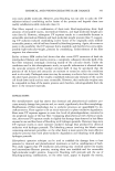
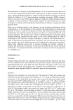
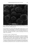
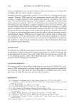
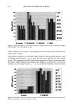
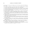
406 JOURNAL OF COSMETIC SCIENCE . : •... /.- . . ,• -.: ß .,,,.. ,.. ß -• ........ jl "•' :" ' 'i: • . . :j •... :.. x . '- .• .-• . . . ,. ,.f . .:.. ...z - ß ':' *'? .•. 't :,- ..•. .. . . . . .. .':, 'r.' ...---- • .. ! ..,,L',' '- • "• . .., :.. . Figure 8. TEM image of isolated hair melanosomes after 120 min of ammonia treatment at pH 10 (x75,000). A suspension of 0.05 mg/ml of hair melanosomes was treated with 1.0% ammonia at pH 10.0. materials. Since the isolation of melanosomes involved sequential treatments with dif- ferent protease enzymes, this amorphous material could not be ordinary proteins, but, rather, highly resistant glycolipids or glycoproteins. A report by Swift (19) indicates a closer association between melanin granules and the cell membrane complex in matured hair samples. Thus, the amorphous material seen in isolated melanosomes could possibly be the cell membrane complex. More work needs to be done to identify the nature of the waxy material surrounding the melanosomes. Sepia eumelanin aggregates are larger (2-5 pm) particles with a "doughnut" shape comprised of 100-150-nm spherical particles, and do not show the waxy material. The ESEM data on Sepia melanin correlates very well to the published SEM data by Nofsinger et al. (11). Magnification of the micron-size particles of Sepia melanosomes showed smaller particles of 100-150 nm in size, indicating that the bigger spherical particles are aggregates of smaller nanoparticles. The reported literature also confirms that the self- assembly of Sepia melanin is a hierarchical process (12). Similar subunits were not seen in isolated hair melanosomes under identical magnification, probably due to the fact that hair melanin nanoparticles are tightly encapsulated in a membrane sac that acts as a protective barrier. Time-dependent TEM imaging studies of ammonia-treated hair melanosomes help us to understand the early events occurring in the bleaching process of isolated hair melano-

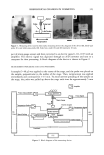
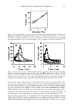
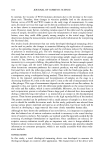
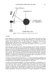
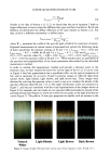
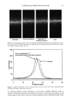
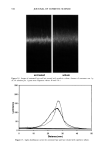
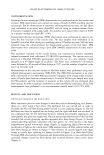
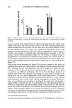
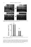
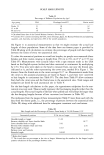
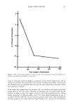
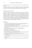
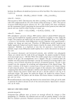
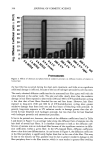
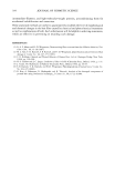
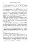
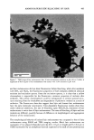
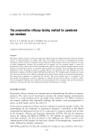
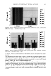
AMMONIA/PEROXIDE BLEACHING OF HAIR 407 j' . 500' nw, Figure 9. TEM image of hair melanosomes after 15 min of treatment with NH3/H202 at pH 10.0 (x40,000). A suspension of 0.05 mg/ml of hair melanosomes was treated with 1.0% ammonia and 1.0% hydrogen peroxide at pH 10.0. somes and provide new insights into the role of ammonia in the bleaching process. Aqueous ammonia treatment results in an initial breakdown of melanosome aggregates by removal of the surrounding amorphous material. This is followed by rupture of the melanosomal membrane sac, releasing melanin nanoparticles (Figure 7). The observation of melanin nanoparticles after ammonia treatment suggests that the micron-size hair melanosomes are comprised of 30-50-nm-size particles. In addition, these studies also provide evidence that, unlike Sepia, hair melanin particles are encapsulated in a mem- brane sac. Prolonged treatment of hair melanosomes with aqueous ammonia induced a complete destruction of characteristic melanosome morphology and a shrinking of the granule size, resulting in an amorphous material (Figure 8). By contrast, Sepia melano- somes under identical conditions of ammonia treatment did not show such changes. This might be due to differences such as metal content and the nature and type of metals present or differences in surface properties and aggregation behavior. Other alkalizing agents such as sodium hydroxide or sodium carbonate at pH 10 did not induce changes to melanosomes similar to those from aqueous ammonia. Thus, it is clear that ammonia-
Purchased for the exclusive use of nofirst nolast (unknown)
From: SCC Media Library & Resource Center (library.scconline.org)