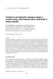
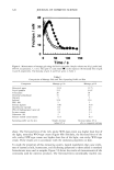
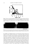
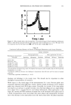
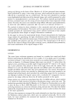
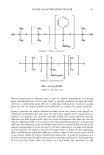
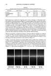
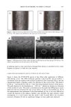
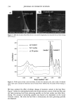
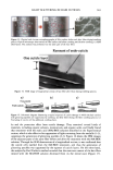
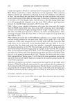
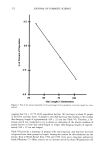
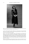
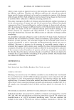
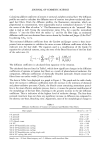
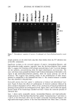
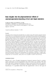
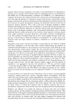
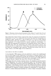
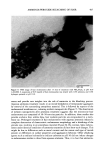
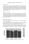
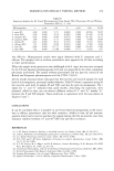
404 JOURNAL OF COSMETIC SCIENCE ß 904.47 100 nm Figure 6. TEM image of isolated hair melanosomes from Asian black hair (x75,000). A sonicated suspen- sion of hair melanosomes (0.05 mg/ml) in water at neutral pH was used for the TEM imaging study. to oxidative attack by hydrogen peroxide. Thus, it is clear from the study that the role of ammonia is to help the release of melanin nanoparticles from the melanosomal sac, making it more susceptible to attack by peroxide. At the end of 30 min, the ammonia/ peroxide-treated melanosome suspension gave a colorless solution devoid of any particles, while ammonia treatment gave a yellow-colored suspension, clearly indicating that the role of peroxide is to decolorize the degraded melanosomes. DISCUSSION Chemically unaltered melanosomes from Asian black hair were isolated using a mild enzymatic procedure involving sequential treatment of a hair sample with different protease enzymes (1). The UV-Vis absorption spectrum of hair melanosome suspension in water showed a structureless spectrum with absorptivity increasing monotonically with decreasing wavelength, characteristic of natural and synthetic melanins. Both Sepia

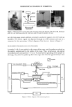
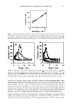
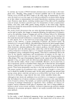
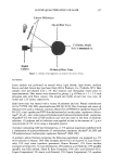
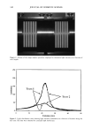
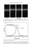
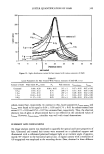
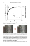
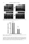
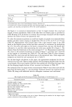
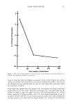
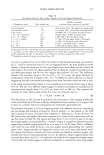
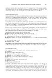
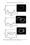
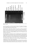
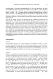
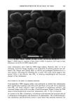
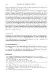
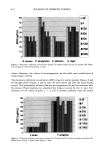
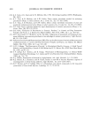
AMMONIA/PEROXIDE BLEACHING OF HAIR 405 Figure 7. TEM image of hair melanosomes after 30 rain of ammonia treatment at pH 10.0 (x75,000). A suspension of 0.05 mg/ml of hair melanosomes was treated with 1.0% ammonia at pH 10.0. and hair melanosomes did not show fluorescence before bleaching, while after oxidation with NH 3 and H202, the fluorescence properties of both melanins exhibited identical emission and excitation spectra. From the excitation spectra, it is clear that a yellow chromophore is responsible for the fluorescence emission properties of melanin after oxidation. This yellow choromophore could be indoquinone or similar quinoid struc- tures resulting from the breakdown and degradation of polymeric melanin as a result of oxidation. The fluorescence data also suggest that Sepia and human hair melanosomes have the same molecular constituents. Time-dependent fluorescence studies show that, under identical conditions, the rate of bleaching upon NH3/H20 2 treatment of hair melanosomes is twice that of Sepia melanosomes. The rates of bleaching of Sepia and hair melanin are different, possibly because of differences in morphological and aggregation behavior of the melanosomes. The morphological behavior of isolated hair melanosomes was compared to that of Sepia melanosomes using ESEM and TEM imaging studies. Black hair melanosomes are aggregates of rice-shaped ellipsoidal particles (0.8-1.0 torn in length and 0.2-0.6 tom in width) surrounded by an amorphous material suspected to be made of non-proteinacious
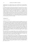
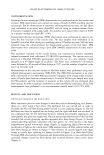
Purchased for the exclusive use of nofirst nolast (unknown)
From: SCC Media Library & Resource Center (library.scconline.org)