CMC STRUCTURE IN HAIR CUTICLE 17
tively short, and our preliminary experiment shows that dye binding for that amount of
time was approximate! y 2 5 %to saturated level.
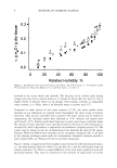
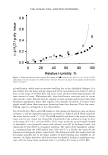
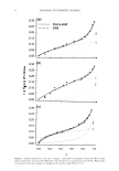
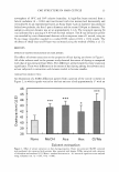
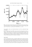
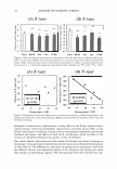
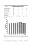
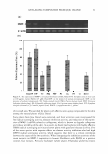
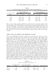
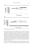
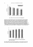
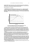
Our findings showed a correlation between the extent of dyeing and the thickness of the
a-layer, which was changed by extraction with the solvents, with a larger decrease in
thickness resulting in a greater elevation in the extent of dyeing. It has been speculated
that hydrophilic molecules penetrate hair through the o-layer, based on histochemical
observations of the CMC (10). Since the dye used in our study (acid orange 7) was
water-soluble, the relationship seen between the extent of dyeing and o-layer thickness
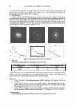
is in agreement with that proposal. Thus, using a microbeam SAXS method, we were
able to detect changes in the CMC structure that correlated with the penetration of
molecules.
CONCLUSION
Microbeam SAXS is a useful tool for hair and cosmetic science. This provides structural
information regarding the cuticular CMC, without the pre-staining or slicing of hair
samples. Using microbeam SAXS, we found CMC structural changes caused by solvent
extraction correlating with changes in the penetration of molecules into the hair. Thus,
using a microbeam SAXS method, we were able to detect changes in the CMC structure
that correlated with the penetration of molecules.
ACKNOWLEDGMENTS
The synchrotron radiation experiments were performed at the SPring-8 facility with the
approval of the Japan Synchrotron Radiation Research Institute QASRI) (Proposal No.
2004B0485-NL2-np).
REFERENCES
(1) J. A. Swift, Human hair cuticle: Biology conspired to the owner's advantage,]. Cosmet. Sci., 50, 23-47
(1999).
(2) C. Robbins, H.-D. Weigmann, S. Ruetsch, and Y. Karnath, Failure of intercellular adhesion in hair
fibers with regard to hair condition and strain conditions,]. Cosmet. Sci., 55, 351-371 (2004).
(3) P.R. Brady, Diffusion of dyes in natural fibres, Rev. Prog. Coloration, 22, 58-78 (1992).
(4) J. D. Leeder, J. A. Rippon, F. E. Rothery, and I. W. Stapleton, Use of transmission electron microscope
to study dyeing and diffusion processes. Proc. 7th Int. Wool Text. Res. Conj Tokyo, 5, 99-108 (1985).
(5) C. L. Gummer, Elucidating penetration pathways into the hair fiber using novel microscopic tech-
niques,]. Cosmet. Sci., 52, 265-280 (2001).
(6) L. Kreplak, C. Merigoux, F. Briki, D. Flot, and]. Doucet, Investigation of human hair cuticle structure
by microdiffraction: Direct observation of cell membrane complex swelling, Biochim. Biophys. Acta,
1547, 268-274 (2001).
(7) N. Ohta, T. Oka, K. Inoue, N. Yagi, S. Kato, and I. Hatta, Structural analysis of cell membrane
complex of a hair fibre by micro-beam X-ray diffraction,]. Appl. Cryst., 38, 274-279 (2005).
(8) K. Inoue, T. Oka, T. Suzuki, N. Yagi, K. Takeshita, S. Goto, and T. Ishikawa, Present status of high
flux beamline (BL40XU) at SPring-8, Nucl. Instrum. Methods Phys. Res. A, 467-468, 674-677 (2001).
(9) K. Joko, J. Koga, and N. Kuroki, The interaction of dyes and wool keratin: The effect of solvent
treatment on dyeing behavior, Proc. 7th Int. Wool Text. Res. Conj, Tokyo, 5, 23-32 (1985).
(10) S. Naito, 1. Takahashi, M. Hatrori, and K. Arai, Hi�i,udH':fri.i.ca.l obsc,,a.tiuu of cell rnernb:-::.!:le CGrr.pleY.:
of hair,]. Soc. Fiber Sci. Tech. Jpn., 48, 420-426 (1992).
tively short, and our preliminary experiment shows that dye binding for that amount of
time was approximate! y 2 5 %to saturated level.
Our findings showed a correlation between the extent of dyeing and the thickness of the
a-layer, which was changed by extraction with the solvents, with a larger decrease in
thickness resulting in a greater elevation in the extent of dyeing. It has been speculated
that hydrophilic molecules penetrate hair through the o-layer, based on histochemical
observations of the CMC (10). Since the dye used in our study (acid orange 7) was
water-soluble, the relationship seen between the extent of dyeing and o-layer thickness
is in agreement with that proposal. Thus, using a microbeam SAXS method, we were
able to detect changes in the CMC structure that correlated with the penetration of
molecules.
CONCLUSION
Microbeam SAXS is a useful tool for hair and cosmetic science. This provides structural
information regarding the cuticular CMC, without the pre-staining or slicing of hair
samples. Using microbeam SAXS, we found CMC structural changes caused by solvent
extraction correlating with changes in the penetration of molecules into the hair. Thus,
using a microbeam SAXS method, we were able to detect changes in the CMC structure
that correlated with the penetration of molecules.
ACKNOWLEDGMENTS
The synchrotron radiation experiments were performed at the SPring-8 facility with the
approval of the Japan Synchrotron Radiation Research Institute QASRI) (Proposal No.
2004B0485-NL2-np).
REFERENCES
(1) J. A. Swift, Human hair cuticle: Biology conspired to the owner's advantage,]. Cosmet. Sci., 50, 23-47
(1999).
(2) C. Robbins, H.-D. Weigmann, S. Ruetsch, and Y. Karnath, Failure of intercellular adhesion in hair
fibers with regard to hair condition and strain conditions,]. Cosmet. Sci., 55, 351-371 (2004).
(3) P.R. Brady, Diffusion of dyes in natural fibres, Rev. Prog. Coloration, 22, 58-78 (1992).
(4) J. D. Leeder, J. A. Rippon, F. E. Rothery, and I. W. Stapleton, Use of transmission electron microscope
to study dyeing and diffusion processes. Proc. 7th Int. Wool Text. Res. Conj Tokyo, 5, 99-108 (1985).
(5) C. L. Gummer, Elucidating penetration pathways into the hair fiber using novel microscopic tech-
niques,]. Cosmet. Sci., 52, 265-280 (2001).
(6) L. Kreplak, C. Merigoux, F. Briki, D. Flot, and]. Doucet, Investigation of human hair cuticle structure
by microdiffraction: Direct observation of cell membrane complex swelling, Biochim. Biophys. Acta,
1547, 268-274 (2001).
(7) N. Ohta, T. Oka, K. Inoue, N. Yagi, S. Kato, and I. Hatta, Structural analysis of cell membrane
complex of a hair fibre by micro-beam X-ray diffraction,]. Appl. Cryst., 38, 274-279 (2005).
(8) K. Inoue, T. Oka, T. Suzuki, N. Yagi, K. Takeshita, S. Goto, and T. Ishikawa, Present status of high
flux beamline (BL40XU) at SPring-8, Nucl. Instrum. Methods Phys. Res. A, 467-468, 674-677 (2001).
(9) K. Joko, J. Koga, and N. Kuroki, The interaction of dyes and wool keratin: The effect of solvent
treatment on dyeing behavior, Proc. 7th Int. Wool Text. Res. Conj, Tokyo, 5, 23-32 (1985).
(10) S. Naito, 1. Takahashi, M. Hatrori, and K. Arai, Hi�i,udH':fri.i.ca.l obsc,,a.tiuu of cell rnernb:-::.!:le CGrr.pleY.:
of hair,]. Soc. Fiber Sci. Tech. Jpn., 48, 420-426 (1992).