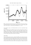
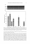
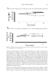
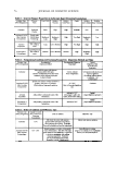
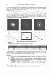
22 JOURNAL OF COSMETIC SCIENCE
N-Succinyl-Ala-Ala-Ala-p-nitroanilide (1.015 mM) was prepared in 0.1232 M Tris-Cl
buffer (pH 8.0) This solution (1300 µl) was added to the sample stock (100 µl). Each
sample stock solution was diluted to final concentrations of 100, 50, and 10 µg/ml. The
solutions were then vortexed and preincubated for 10 min at 25°C, and elastase (0.0375
unit/ml) stock solution (100 µl) was added. After vortexing, the solutions were placed
in a water bath for 10 min at 25°C. The absorbance was measured at 410 nm.
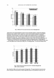
CYTOTOXICITY ASSAY ON HUMAN FIBROBLASTS
Human fibroblast cells (American Type Culture Collection, ATCC, CRL-2076) were
seeded in 24-well plates with DMEM +10% FBS at a density of 1 x 10⁵ cells per well
and cultured at 37°C in 5% CO2 .After one day, the medium was exchanged with fresh
media containing 2% serum, and the cells were allowed to incubate in a CO2 incubator
at 37°C in the presence of samples (100 µ1/ml) for 24 h. The cells were then treated with
100 µl of 2.5 mg/ml MTT and incubated at 37°C for an additional 4 h. The medium
containing the MTT was discarded, the MTT formazan produced was extracted with 1
ml of DMSO, and the absorbance was read at a wavelength of 5 70 nm with a reference
wavelength of 650 nm. The level of cell viability was calculated as:
Cell viability(%)=(OD
57 o
(
sample/OD570
(comrol ))X 100
where OD 57 o(sample) is the absorbance of the treated cells at 570 nm and OD
57 o(control)
is the absorbance of the negative control (non-treated cells) at 5 70 nm.
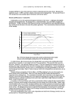
INHIBITION ASSAY ON MMP-1 EXPRESSION BY RT-PCR
Human fibroblasts were cultured with DMEM +10% FBS, 50 U/mol penicillin, and 50
µg/ml of streptomycin the medium was changed every two or three days. Cells were
cultured at 3 7 ° C in 5 %CO2 • When the cells reached confluence, they were separated
by treatment with 0.25% trypsin-0.03% EDTA (ethylenediamine tetraacetic acid) so-
lution. Cells were seeded into a 100-mm dish at a density of 2 x 10⁶ cells and cultured
at 37°C in 5% CO2. After one day, fresh medium containing 2% serum was added to
the cells, which were then treated with samples for 24 hours. Total RNA was isolated
from the cells with TRizol (Invitrogen) according to the instructions of the manufac-
turer. First-strand cDNA synthesis was performed using random hexamers. The se-
quences of primers are as follows: 5 '-TGGGAGCAAACACATCTGA-3' (sense) and
5'-ATCACTTCTCCCCGAATCGT-3' (anti-sense) for MMP-1 5'-GAGACCTTCA-
ACACCCCAGCC-3' (sense) and 5'-GGCCATCTCTTGCTCGAAGTC-3' (anti-sense)
for �-actin. For MMP-1 RT-PCR reactions, reverse transcription was performed at 50°C
for 30 min, and denaturing was performed at 96°C for 3 min, followed by 22 cycles at
94°C for 1 min, 48°C for 1 min, and 72°C for 1 min, followed by an extension step cycle
at 72°C for 10 min. For �-Actin RT-PCR reactions, reverse transcription was performed
at 50°C for 30 min, and denaturing was performed at 96°C for 3 min, followed by 29
cycles at 94°C for 1 min, 70°C for 1 min, and 72°C for 1 min, followed by an extension
step cycle at 72°C for 10 min. The final products were detected with 1.5% agarose gel.
The gels were photographed, and the intensity of the stained PCR fragments was
quantified from photographs by densitometric analysis using Gel Doc 2000 (Bio-Rad
Laboratories, Segrate, Milan, Italy). EGCG ((-)epigallocatechin-3-gallate) was used as a
positive control.
N-Succinyl-Ala-Ala-Ala-p-nitroanilide (1.015 mM) was prepared in 0.1232 M Tris-Cl
buffer (pH 8.0) This solution (1300 µl) was added to the sample stock (100 µl). Each
sample stock solution was diluted to final concentrations of 100, 50, and 10 µg/ml. The
solutions were then vortexed and preincubated for 10 min at 25°C, and elastase (0.0375
unit/ml) stock solution (100 µl) was added. After vortexing, the solutions were placed
in a water bath for 10 min at 25°C. The absorbance was measured at 410 nm.
CYTOTOXICITY ASSAY ON HUMAN FIBROBLASTS
Human fibroblast cells (American Type Culture Collection, ATCC, CRL-2076) were
seeded in 24-well plates with DMEM +10% FBS at a density of 1 x 10⁵ cells per well
and cultured at 37°C in 5% CO2 .After one day, the medium was exchanged with fresh
media containing 2% serum, and the cells were allowed to incubate in a CO2 incubator
at 37°C in the presence of samples (100 µ1/ml) for 24 h. The cells were then treated with
100 µl of 2.5 mg/ml MTT and incubated at 37°C for an additional 4 h. The medium
containing the MTT was discarded, the MTT formazan produced was extracted with 1
ml of DMSO, and the absorbance was read at a wavelength of 5 70 nm with a reference
wavelength of 650 nm. The level of cell viability was calculated as:
Cell viability(%)=(OD
57 o
(
sample/OD570
(comrol ))X 100
where OD 57 o(sample) is the absorbance of the treated cells at 570 nm and OD
57 o(control)
is the absorbance of the negative control (non-treated cells) at 5 70 nm.
INHIBITION ASSAY ON MMP-1 EXPRESSION BY RT-PCR
Human fibroblasts were cultured with DMEM +10% FBS, 50 U/mol penicillin, and 50
µg/ml of streptomycin the medium was changed every two or three days. Cells were
cultured at 3 7 ° C in 5 %CO2 • When the cells reached confluence, they were separated
by treatment with 0.25% trypsin-0.03% EDTA (ethylenediamine tetraacetic acid) so-
lution. Cells were seeded into a 100-mm dish at a density of 2 x 10⁶ cells and cultured
at 37°C in 5% CO2. After one day, fresh medium containing 2% serum was added to
the cells, which were then treated with samples for 24 hours. Total RNA was isolated
from the cells with TRizol (Invitrogen) according to the instructions of the manufac-
turer. First-strand cDNA synthesis was performed using random hexamers. The se-
quences of primers are as follows: 5 '-TGGGAGCAAACACATCTGA-3' (sense) and
5'-ATCACTTCTCCCCGAATCGT-3' (anti-sense) for MMP-1 5'-GAGACCTTCA-
ACACCCCAGCC-3' (sense) and 5'-GGCCATCTCTTGCTCGAAGTC-3' (anti-sense)
for �-actin. For MMP-1 RT-PCR reactions, reverse transcription was performed at 50°C
for 30 min, and denaturing was performed at 96°C for 3 min, followed by 22 cycles at
94°C for 1 min, 48°C for 1 min, and 72°C for 1 min, followed by an extension step cycle
at 72°C for 10 min. For �-Actin RT-PCR reactions, reverse transcription was performed
at 50°C for 30 min, and denaturing was performed at 96°C for 3 min, followed by 29
cycles at 94°C for 1 min, 70°C for 1 min, and 72°C for 1 min, followed by an extension
step cycle at 72°C for 10 min. The final products were detected with 1.5% agarose gel.
The gels were photographed, and the intensity of the stained PCR fragments was
quantified from photographs by densitometric analysis using Gel Doc 2000 (Bio-Rad
Laboratories, Segrate, Milan, Italy). EGCG ((-)epigallocatechin-3-gallate) was used as a
positive control.