196 JOURNAL OF COSMETIC SCIENCE cells to injury it may also be involved in the deposition of connective tissue matrix proteins in diverse states such as angiogenesis and organogenesis, as well as in patho logical fibrotic states (5 ,6). TGF-J3 l induces collagen synthesis and has been shown to be upregulated in scars. In addition, TGF-J3 l has been shown to promote the growth of human fibroblasts into stratified layers, mimicking in vivo fibroplasia (7 ,8). Previous reports have described the anti-melanogenic effects of placental extract however, in a Medline search of the literature published between 195 7 and 2006, we found no studies evaluating the effects of placental extract on fibroblast proliferation associated with collagen synthesis. We investigated the effects of placental extract on fibroblast proliferation and TGF-J3 l expression. To validate our findings, we performed experiments on both placental extract and ascorbic acid, which has been shown to affect fibroblast proliferation. Placental extract increased fibroblast proliferation but did not significantly increase TGF-J31 expression compared to the controls. MATERIALS AND METHODS HUMAN SKIN FIBROBLAST CULTURE Primary cultures of normal human skin fibroblasts were established from newborn prepuce in Dulbecco's modified Eagle's medium (DMEM) supplemented with 10% fetal bovine serum (FBS), glutamine 2 mM, penicillin 100 U/ml, and streptomycin 100 µg/ml at 37 ° C in a humidified incubator containing 5% CO2 . The fibroblasts were cultured to 90% confluence and then subcultured. For all assays, fibroblasts at fourth passages were used. TREATMENT OF FIBROBLASTS WITH PLACENTAL EXTRACTS AND ASCORBIC ACID Cultured human fibroblasts were treated with placental extracts (Melsmon Pharmaceu tical Co. Ltd., Tokyo, Japan) and ascorbic acid (L-ascorbic acid-2-phosphate magnesium Sigma, St. Louis, MO). We stored the placental extract under temperatures between 2° and 8°C, and carried out the experiments as soon as the extract was received from the manufacturer, although the valid period is two years according to the instructions. The fibroblasts were exposed to various concentrations of placental extract (0, 0.08, 0.16, 0.32, and 0.64%) and ascorbic acid (0, 0.01, 0.10, 1, and 10 mM). We regarded cultured human fibroblasts without any treatment as the control. All experiments were performed independently three times. CELL PROLIFERATION ASSAY Cell proliferation was determined by MTT assay. A CellTiter 96 aqueous proliferation assay kit (Promega, Madison, WI) was used for the cell viability test in accordance with the manufacturer's instructions. Fibroblasts were seeded in 96-well plates at a density of 1 x 10⁴ cells/well and were incubated for 48 h. The fibroblasts were then treated with placental extract (0.08, 0.16, 0.32, and 0.64%) and ascorbic acid (0.01, 0.1, 1, and 10 mM) and incubated for 24 h. A total of 20 µl of Cell Titer reagent containing tetrazolium

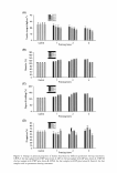
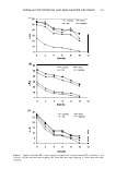
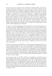
EFFECTS OF PLACENTAL EXTRACT ON FIBROBLAST PROLIFERATION 197 and an electron-coupling reagent was added to each well and catalyzed by mitochondrial dehydrogenase enzymes in metabolically active cells. The plates were incubated for 4 h and the colorimetric absorbance was recorded at 5 7 0 nm using a microplate reader. The results were expressed as a percentage of the controls. MEASURING TGF-131 PROTEIN EXPRESSION BY ENZYME-LINKED IMMUNOSORBENT ASSAY (ELISA) Fibroblasts were seeded at a density of 1 x 10⁶ cells/ml per 100-mm dish and cultured for 48 h. The culture medium was replaced by DMEM without FBS three days before the experiment. The fibroblasts were then treated with different concentrations of pla cental extract and ascorbic acid for 24 h. Culture supernatants were collected, centri fuged at 1000 x g for 5 min, and stored at -80°C until an ELISA was performed for TGF-131. The volumes of culture supernatants were adjusted to 1 x 10⁶ cells/ml. TGF-131 protein was quantified using a total human TGF-131 ELISA kit (R&D Systems, Minneapolis, MN). Briefly, the ELISA plates were coated with 100 µl of antihuman TGF-131 antibodies that had been diluted in 1 x Voller's buffer (pH 9.6) and stored at 4°C overnight. Test samples were activated with 1 N HCl for 10 min and neutralized with 1.2 N NaOH/0.5 M HEPES at room temperature. The plates were washed, and the samples were added in duplicate to individual wells and incubated at room temperature for 2 h. After three washes, 100 µl of biotinylated antibodies diluted in PBS (pH 7.4) containing 0.05% Tween 20 was added for 1 h. After washing, 100 µl of streptavidin horseradish peroxidase (HRP) conjugate that had been diluted to 1 :20,000 in a dilution buffer was added for 1 h. After a final wash, 200 µl of the HRP substrate tetrameth ylbenzidine dihydrochloride and hydrogen peroxide in 0.05 M phosphate-citrate buffer (pH 5.0) were added for 30 to 60 min. The reaction was stopped by adding 50 µl of 1 M sulfuric acid, and the absorbence at 450 nm was determined with an EMax microplate reader (Molecular Devices, Sunnyvale, CA). Protein levels were determined by compar ing the absorbences produced by the test samples versus those produced by the stan dards. Existing levels of TGF-131 in placental extract were excluded in the measures of the placental extract-treated groups. STATISTICAL ANALYSIS Statistical analyses were performed using the Statistical Package for Social Sciences version 12.0 (SPSS Inc., Chicago, IL). Student's two-tailed t-test was used to evaluate the differences between the study groups. P values less than 0.05 were considered statisti cally significant. RESULTS EFFECTS OF PLACENTAL EXTRACT AND ASCORBIC ACID ON FIBROBLAST PROLIFERATION To clarify the effects of placental extract on fibroblast proliferation, fibroblasts were treated with placental extract at concentrations of 0, 0.08, 0.16, 0.32, and 0.64% and ascorbic acid at concentrations of 0, 0.01, 0.1, 1.0, and 10 mM. Placental extract concentrations of 0.08 and 0.16% (% of controls, 103.4 ± 5.9% and 104.9 ± 3.4%, respectively) did not show a significant effect on fibroblast proliferation compared to the
Purchased for the exclusive use of nofirst nolast (unknown)
From: SCC Media Library & Resource Center (library.scconline.org)