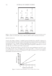
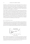
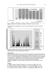
JOURNAL OF COSMETIC SCIENCE 478 was transferred to the wells of a black 96-well plate. The samples and standards were as- sayed in duplicate. After the samples/standards were added to the plate, 100 μl of the diluted Ribogreen assay reagent was added to the wells, and the plate was gently mixed and allowed to incubate for 5–10 minutes protected from light. After incubation, the plate was read by a fl uorometer using an excitation wavelength of 500 nm and an emis- sion wavelength of 525 nm. mRNA AMPLIFICATION (Ambion MessageAmp aRNA Kit) First-strand cDNA synthesis. Five micrograms of total RNA for each sample was placed into 600-μl PCR tubes, and the total volume of liquid in each tube was adjusted to 12 μl with DEPC·H2O. To each tube, 1 μl of T7 Oligo(dT) primer was added, and the tube was incubated at 70 ± 2°C for 10 minutes to denature the RNA. The sample was then placed onto ice to allow the primer to anneal to the poly A ends of the mRNA. After cooling, 2 μl of 10X fi rst-strand buffer, 1 μl of RNAse inhibitor, and 4 μl of dNTP mix was added to each tube and the tubes were placed at 42 ± 2°C. As soon as the tube was heated, 1 μl of reverse transcriptase was added and the tubes were returned to the 42 ± 22°C bath for two hours. After heating, the tubes were briefl y centrifuged to gather the fl uids at the bottom of the tube, and then they were cooled on ice. Second-strand synthesis and cDNA amplifi cation. For the synthesis of the second strand of cDNA, the following items were added to the tubes described above in the following order: 63 μl of DEPC·H2O, 10 μl of 10X second-strand buffer, 4 μl of dNPT mix, 2 μl of DNA polymerase, and 1 μl of RNAse H. Each tube was mixed and then incubated at 16 ± 2°C for two hours. Toward the end of the two-hour incubation, a suffi cient quantity of DEPC·H2O was added, the tube was warmed to 50 ± 2°C, and a cDNA purifi cation fi lter cartridge was equilibrated with 50 μl of cDNA binding buffer (one cartridge per sample) for at least fi ve minutes. After the samples had fi nished incubating, 250 μl of cDNA binding buffer was added to each tube and the tubes were thoroughly mixed. The contents of the each PCR tube were transferred to the cDNA purifi cation fi lter cartridge. The cartridge was then placed in a collection tube and centrifuged at 10,000 rpm for one minute. The fl ow-through was discarded and 650 μl of cDNA wash solution was added to the cartridge. The cartridge was centrifuged again and the fl ow-through was discarded. The contents were centrifuged one more time to ensure that the wash buffer was completely emptied from the fi lter. The cDNA was eluted by applying 10 μl of preheated DEPC·H2O to the fi lter, and the contents were centrifuged in a new collection tube at 10,000 rpm for one minute. The elution was repeated one additional time to give a total volume of 16–18 μl of cDNA. In vitro transcription to synthesize aRNA and aRNA purifi cation. The in vitro transcription began by adding the following to the cDNA solution: 4 μl of 10X reaction buffer and 4 μl of T7 enzyme mix. The tube was mixed and then incubated at 37 ± 2°C for 6–14 hours. Towards the end of the incubation period, a suffi cient volume of elution solution was warmed to 50°–60°C and an aRNA fi lter cartridge was equilibrated with 100 μl of aRNA binding buffer for at least fi ve minutes. At the end of the incubation period, 350 μl of aRNA binding buffer was added to the sample tubes and thoroughly mixed. An additional 250 μl of absolute ethanol was added to each tube. The mixture was trans- ferred to an aRNA fi lter cartridge, and the cartridge was then inserted into a collection tube and centrifuged at 10,000 rpm for one minute. The fl ow-through was discarded and 650 μl of aRNA wash buffer was added to the cartridge, followed by further centrifugation

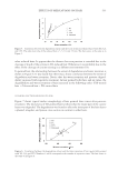
KUDZU EXTRACT AND PROCOLLAGEN PRODUCTION 479 at 10,000 rpm for one minute. After the fl ow-through was discarded, the cartridge was spun one fi nal time to remove traces of the wash buffer. The cartridge was transferred to a new collection tube and 25 μl of prewarmed elution solution was added to the cartridge. The cartridge was incubated for two minutes at room temperature and then aRNA was eluted by centrifuging for one minute at 10,000 rpm. This elution was performed one additional time to give a total volume of 45–50 μl of aRNA solution. The fi nal concentra- tion of the aRNA was determined by the Ribogreen assay described above. In addition, the quality of the aRNA was checked via gel electrophoresis as described below. Labeling of aRNA with fl uorescent dyes (Perkin Elmer ASAP RNA Labeling Kit) and purifi cation of labeled aRNA. Labeling: Two tubes were prepared for the labeling process, one for Cy3 labeling (green) and one for Cy5 labeling (red). To the Cy3 tube was added 2 μg of aRNA prepared from the untreated control sample, and enough DEPC·H2O was added to bring the total vol- ume up to about 4 μl. To the Cy5 tube was added 2 μg of aRNA prepared from the sample treated with the test material, and enough DEPC·H2O was added to bring the total volume up to 4 μl. To both tubes was added 5 μl of ASAP labeling buffer and 1 μl of the specifi c dye for each tube (Cy3 or Cy5). The tubes were incubated for 15 minutes at 85 ± 2°C. At the end of the 15-minute incubation period, the tubes were placed on ice to cool and then 2.5 μl of ASAP stop solution was added to each tube. The proportions provided enough sample to run one microarray chip. Purifi cation: To purify the labeled aRNA, a Microcon YM-30 fi lter column (Millipore) was inserted into a collection tube fi lled with 400 μl of TE buffer. The Cy3 and Cy5 probes were combined (12.5 μl of each) and then added to the Microcon fi lter the mix- ture was thoroughly mixed with the TE buffer. The mixture was centrifuged at 12,000 rpm for eight minutes and the fl ow-through was discarded. The column was then washed twice with 400 μl of TE buffer and the fl ow-through was again discarded. After the fi nal wash, the fi lter column was inverted, placed into a new collection tube, and centrifuged at 12,000 rpm for two minutes to collect the probe. The probe was further diluted to a volume of 2–30 μl with residual TE buffer. Microarray hybridization and washing (Agilent Technologies Microarrays). For hybridization, 45 μl of 10X control target RNA (Agilent Technologies In Situ Hybridization Kit) was mixed with 160 μl of DEPC water and 9 μl of 25X Agilent fragmentation buffer. This mixture was incubated at 60°C for approximately 30 minutes in a hybridization oven. At the end of the incubation period, 225 μl of Agilent hybridization buffer was added, along with the fl uorescent aRNA probes prepared above. During the incubation period, an Agilent SUREHYB hybridization chamber was prepared by inserting a glass gasket slide into the bottom half of the chamber. At the end of the incubation period, the hybridiza- tion mixture (approximately 450 μl) was applied to the glass gasket slide and an Agilent Human 1A Oligo microarray chip was placed face down on top of the gasket in such a way that the hybridization solution was sandwiched between the glass gasket slide and the microarray face of the chip. The top half of the chamber was then attached and the connecting thumbscrew tightened. After verifying that there was good bubble formation in the chamber, it was placed into the hybridization chamber for approximately 17 hours at 60°C and rotated at 4 rpm). At the end of the hybridization period, the microarray glass gasket assembly was removed from the SUREHYB chamber and placed in 50 ml of wash solution 1 (6X SSC, 0.005% Triton X-102) at room temperature. After the gasket
Purchased for the exclusive use of nofirst nolast (unknown)
From: SCC Media Library & Resource Center (library.scconline.org)