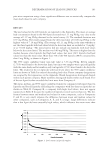
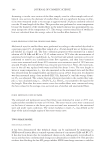
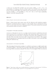
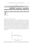
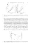
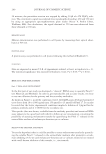
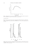
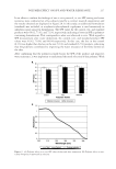
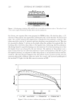
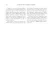
JOURNAL OF COSMETIC SCIENCE 202 molecule, which can polymerize into extracellular collagen fi brils. Thus, the amount of the free propeptides stoichiometrically refl ects the amount of collagen molecules synthe- sized. In a similar manner, we reasoned that the soluble part of Pmel 17 could refl ect the amount of total Pmel 17 and therefore serve to assess melanosome transfer. The fact that a well-known melanosome transfer inhibitor (niacinamide) signifi cantly reduced the amount of soluble Pmel 17 found in the extracellular media of our co- cultures fi ts well with this hypothesis. Moreover, as niacinamide has never been demon- strated to modify the activity of zinc-dependent proteinases (i.e., metalloproteinases, MMP, and Disintegrin-type metalloproteinases, ADAMs), which are the main proteases implicated in the protein ectodomain shedding phenomenon (for a review, see references 9 and 10), we can reasonably conclude that our strategy permits the measurement of me- lanosome transfer in human-cell co-culture models. In the second part of our study, we chose to use our newly developed model to defi ne more precisely the signaling pathways implicated in the whitening effect of a commercially available Alaria esculenta extract (unpublished data). As the algae extract we have selected is able to signifi cantly reduce the activation of PAR-2 (unpublished data), a receptor im- plicated in the melanosome uptake by the keratinocyte, we examined whether it could also act through the melanosome transfer to produce its lightening effects. As shown in Figure 2, the Alaria esculenta extract at 0.01% (v/v) was able to signifi cantly reduce the amount of Pmel 17 found in the co-culture media: −15.4% (p0.05), without affecting melanogenesis (Figure 3). This suggests that the whitening effect of our algae extract is more likely due, at least in part, to its inhibitory activity on the melanosome transfer. The fact that the maximal effect was observed for the lowest tested concentration seems to indicate that side effects were also triggered when higher concentrations were tested. In order to better characterize the effect of the Alaria esculenta extract on the me- lanosome transfer and/or to link this effect to its activity on the PAR-2 receptor, addi- tional experiments should be carried out. Further experiments, using different purifi ed fractions of the Alaria esculenta extract are currently in progress. CONCLUSION For the fi rst time we have provided a simple and effi cient way to quantitatively assay melano- some transfer in human normal melanocyte–keratinocyte co-cultures. Our methodological Figure 3. Effect of Alaria esculenta extract on melanogenesis.

MELANOSOME TRANSFER EVALUATION 203 and technological approach is easily adaptable to the achievement of middle- or high- throughput screenings and could then provide a useful tool for the development of new cosmetic whitening products. It could also be used as a way to confi rm and quantify the whitening action of currently developed products. ACKNOWLEDGMENTS The authors are very thankful to Julie Le Luhant (EFFISCIENCE) for very good technical support. The authors also thank Andrew Menzies (www.menzies.fr) for his rapid and un- derstanding linguistic support. REFERENCES (1) W. Berens, K. Van den Bossche, T. J. Yoon, W. Westbroek, J. C. Valencia, C. J. Out, J. M. Naeyaert, V. J. Hearing, and J. Lambert, Different approaches for assaying melanosome transfer, Pigment Cell Res., 18, 370–381 (2005). (2) S. K. Singh, C. Nizard, R. Kurfurst, F. Bonte, S. Schnebert, and D. J. Tobin, The silver locus product (Silv/gp100/Pmel17) as a new tool for the analysis of melanosome transfer in human melanocyte– keratinocyte co-culture, Exp. Dermatol., 17, 418–426 (2008). (3) T. Hoashi, K. Tamaki, and V. J. Hearing, The secreted form of a melanocyte membrane-bound glyco- protein (Pmel17/gp100) is released by ectodomain shedding, FASEB J., 24, 916–930 (2010). (4) J. F. Berson, D. C. Harper, D. Tenza, G. Raposo, and M. S. Marks, Pmel17 initiates premelanosome morphogenesis within multivesicular bodies. Mol. Biol. Cell, 12, 3451–3464 (2001). (5) T. Hakozaki, L. Minwalla, J. Zhuang, M. Chhoa, A. Matsubara, K. Miyamoto, A. Greatens, G. G. Hillebrand, D. L. Bissett, and R. E. Boissy, The effect of niacinamide on reducing cutaneous pigmenta- tion and suppression of melanosome transfer, Brit. J. Dermatol., 147, 20–31 (2002). (6) A. Greatens, T. Hakozaki, A. Koshoffer, H. Epstein, S. Schwemberger, G. Babcock, D. Bissett, H. Takiwaki, S. Arase, R. R. Wickett, and R. E. Boissy, Effective inhibition of melanosome transfer to keratinocytes by lectins and niacinamide is reversible, Exp. Dermatol., 14, 498–508 (2005). (7) M. M. Bradford, A rapid and sensitive method for the quantitation of microgram quantities of protein utilizing the principle of protein-dye binding, Anal. Biochem., 72, 248–254 (1976). (8) M. B. Taubman, B. Goldberg, and C. Sherr, Radioimmunoassay for human procollagen, Science, 186, 1115–1117 (1974). (9) K. J. Garton, P. J. Gough, and E. W. Raines, Emerging roles for ectodomain shedding in the regulation of infl ammatory responses, J. Leukocyte Biol., 79, 1105–1116 (2006). (10) P. Montes de Oca-B, Ectodomain shedding and regulated intracellular proteolysis in the central nervous system, Cent. Nerv. Syst. Agents Med. Chem., 10, 337–359 (2010).
Purchased for the exclusive use of nofirst nolast (unknown)
From: SCC Media Library & Resource Center (library.scconline.org)