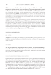
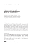
JOURNAL OF COSMETIC SCIENCE 360 Methionines may act like free radical sinks by reacting with ROS to form metSO on the periphery of a protein and thus protect the protein (6). Although methionine is oxidized in the process, the reactive intermediate is disarmed and the overall integrity of the pro- tein is preserved. metSO may then be regenerated back to methionine by methionine sulfoxide reductase A (MSRA) using thioredoxin as a cofactor (7). This strategy may be advantageous to a cell because the time for repair to occur may be a critical factor for cell maintenance or it may be energetically more favorable for the cell to repair itself rather than to resynthesize a new protein. In this study, MSRA levels were evaluated in normal human epidermal keratinocytes (NHEK) before and after UVB exposure. MSRA’s protective effect was also determined by a direct addition of this enzyme to cells before irradiation. Further, NHEK were pre- treated with a metSO-containing pentapeptide (mp) in order to determine if MSRA could be induced in a non-cytotoxic manner and provide an enhanced antioxidant defense mechanism. Lastly, this peptide was added to the media of skin models and tested for its ability to inhibit sunburn cell production after UVB treatment. MATERIALS AND METHODS CELL CULTURE NHEK were obtained from Cascade Biologics (Portland, OR) as primary culture male cells. Cells were maintained in EpiLife (calcium-free) medium containing 1% supplemented serum (Cascade). SKIN MODELS EFT 400 skin models were obtained from MatTek (Ashland, MA) and maintained with a transwell membrane air/liquid interface as per the manufacturer’s instructions. After treatment, histological examination was carried out by hematoxylin and eosin staining (Paragon Bioservices, Baltimore, MD). REVERSE TRANSCRIPTION POLYMERASE CHAIN REACTION Conventional reverse transcription polymerase chain reaction (RT-PCR) was performed on RNA extracts for msrA transcription using primers obtained from Invitrogen (Carlsbad, CA): GTGGTGTTCCAGCCCGAGCACAT (sense) and ATGTCGGTGGTGATCAG GCCGAA (antisense). β-Microglobulin or gapdh was used as housekeeping genes. Fol- lowing annealing, reverse transcription, and amplifi cation, amplicons were separated on 2% agarose gels. Gels were stained with SYBR Gold (Invitrogen) and visualized by UV transillumination. Images were captured with a charge-coupled device (CCD) camera and quantitated with UnScanIt imaging software (Silk Scientifi c, Orem, UT).

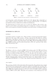
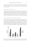
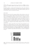
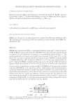
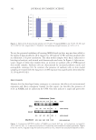
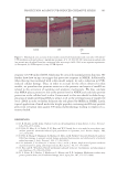
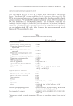
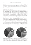
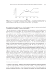
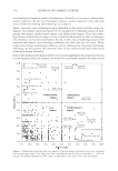
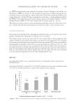
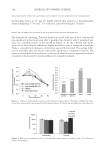
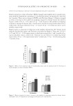
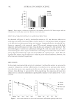
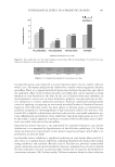
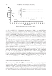
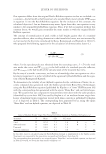
PROTECTION AGAINST UVB-INDUCED OXIDATIVE STRESS 361 HYDROGEN PEROXIDE DETERMINATION Hydrogen peroxide (H2O2) was measured as described previously (8). Briefl y, cells were treated with 10 μM 2′,7′-dichlorodihydrofluorescein diacetate (Molecular Probes, Eugene, OR) for 6 h and their fl uorescence measured (Ex485 nm /Em530 nm ). CELL VIABILITY Cell viability was evaluated by an MTS assay as described previously (9). MSRA AND METHIONINE SULFOXIDE PEPTIDE MSRA was obtained as an experimental test sample from Promega (Madison, WI). The pentapeptide containing metSO was synthesized and obtained from MMP Inc. (Plainfi eld, NJ). RESULTS NHEK were irradiated in Dulbecco’s phosphate-buffered saline, pH 7.4 with 50 mJ/cm2 UVB, the RNA isolated, and subjected to RT-PCR. The results from this analysis showed an increase in msrA expression in response to environmental trauma (Figure 1). The gapdh-normalized msrA levels (msrA/gapdh ratio) increased 22.6% from 0.53 to 0.65 (±0.07 S.E.). In order to establish whether increased amounts of MSRA could reduce ROS in NHEK following UVB irradiation, NHEK (2 × 10⁴) were pre-treated with 0.35 and 0.7 mg/ml of MSRA overnight and then exposed to 20 mJ/cm2 UVB. As shown in Figure 2, H2O2 decreased by 24.7% (±3.3% S.E.) and 36.1% (±0.5% S.E.), respectively. In a next series of experiments, an mp was added to NHEK for 24 h, harvested, and then analyzed by RT-PCR for msrA expression as before. Additionally, an identical pentapeptide containing methionine instead of metSO was tested in parallel. The results from these experiments demonstrated an 18.2% (±4.0% S.E.) increase in msrA expression in the cells treated with only 0.01 mg/ml mp, whereas a similar increase was not observed in the met-containing mp (Figure 3). Increases were observed at the 0.1 and 0.25 mg/ml con- centrations as well. Further, no cytotoxicity was measured for either pentapeptide as deter- mined by the MTS assay for cell viability (data not shown). Figure 1. Conventional RT-PCR analysis of UVB-irradiated NHEK showed an average increase in msrA expression when normalized to gapdh (0.53 vs. 0.65 ± 0.07 S.E.).
Purchased for the exclusive use of nofirst nolast (unknown)
From: SCC Media Library & Resource Center (library.scconline.org)