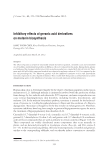
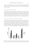
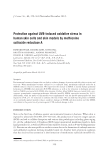
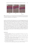
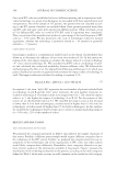
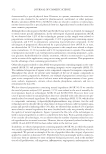
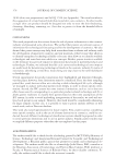
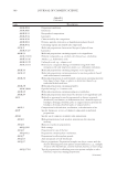
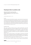
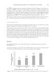
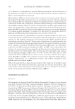
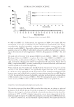
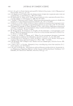
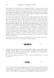
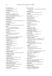
JOURNAL OF COSMETIC SCIENCE 352 acid and geranic acid by subsequent oxidation (14–16). Among these compounds, ge- ranic acid has recently been reported to exert a tyrosinase-inhibitory effect (11). However, no study has yet been conducted on its effects on melanocytes. The principal objectives of this study were to evaluate the inhibitory effects of these com- pounds on cell viability and melanin production in melanocytes, and also to assess their effects on the expression of melanin biosynthesis-associated enzymes, including tyrosinase. EXPERIMENTAL METHODS MATERIALS Citronellol, geraniol, citronellic acid, L -Dopa, and kojic acid were purchased from Sigma- Aldrich Co. (St. Louis, MO). Geranic acid was acquired from the Fluka Co. (Buchs, Switzerland). The Melan-a cell line was a gift from Dr. Bennett (St. George’s Hospital Medical School, London, UK). Fetal bovine serum (FBS), Roswell Park Memorial Insti- tute (RPMI) medium, and Penicillin–Streptomycin (PS) were purchased from Gibco BRL. (Grand Island, NY) CELL CULTURE PROCEDURES The Melan-a cells were cultured in RPMI 1640 medium under 10% FBS and 200-nm phorbol 12-myristate 13-acetate (PMA) conditions. In 100 ml culture dishes, 10 ml of medium was added and then seeded with 5 × 10⁵ cells. The cells were grown to confl u- ence after 3 to 4 days at 37°C in an atmosphere of 5% CO2, they were seeded with 105 cells/well in a 24-well plate, and then incubated for 24 h. Each well was replenished with 990 μl of medium daily, as well as treated with 10 μl of test sample [solvent (v/v): pro- pylene glycol/EtOH/H2O = 5/3/2] for 3 days the plate was then incubated for 1 day (17). CELL VIABILITY The percentage of viable cells was determined by staining the cell population using a simple crystal violet (CV) staining method to quantitate adherent cell number after treatment (18). After the removal of media from each well, the wells were washed with phosphate-buffered saline (PBS). Two hundred microliters of CV (0.1% CV, 10% EtOH, and the remaining volume as PBS) was then added. The plates were incubated at room temperature for 5 min Figure 1. Chemical structure of geranic acid and its derivatives.

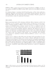
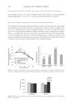
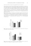
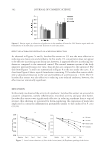
INHIBITORY EFFECTS OF GERANIC ACID 353 and washed twice with water. After the addition of 1 ml of EtOH, the plates were shaken for 10 min at room temperature. UV absorption was measured at 590 nm (19). DETERMINING MELANIN CONTENT After the removal of media from each well, the plate was washed with PBS, followed by the addition of 1 ml of 1 N NaOH to each well to lyse the cells for the release of melanin. The UV absorption was measured at 405 nm. Phenylthiourea (PTU) was employed as a positive control (20). WESTERN IMMUNOBLOTTING ANALYSIS The Melan-a cells were harvested and extracted in a triple-detergent lysis buffer [50 mM Tris-HCl (pH 8.0), 150 mM NaCl, 0.02% sodium azide, 0.1% sodium dodecyl sulfate (SDS), 1% nonyl phenoxypolyethoxylethanol (NP-40), 0.5% sodium deoxycholate, 100 μg/ml of phenylmethylsulfonyl fl uoride (PMSF), and 1 μg/ml of aprotinin]. The protein content was then measured with a protein assay kit (Bio-rad, Hercules, CA). Next, 50 μg of the protein was separated on 8% SDS-polyacrylamide gel and transferred to a Hybond enhanced chemiluminescence (ECL) nitrocellulose membrane (Amersham Pharmacia Biotech, Buckinghamshire, UK). The membranes were blocked with 5% skim milk and incubated with tyrosinase (Santa Cruz Biotech, Santa Cruz, CA 1/250 dilution) primary antibody or dopachrome tautomerase (TRP-2, Santa Cruz Biotech, 1/300 dilution) pri- mary antibody, and anti-goat secondary antibodies. Detection was performed using ECL (Amersham Pharmacia Biotech, Piscataway, NJ). The Western blot results obtained by the scanner were photographed (HP, Palo Alto, CA). Density of protein bands was mea- sured using Image J program by National Institute of Health (NIH) (21). MEASURING INHIBITORY EFFECT ON TYROSINASE ACTIVITY Tyrosinase activity was measured by its dopa-oxidase activity, using a slightly modifi ed version of the method reported by Shono and Toda (22). Each concentration (1 mM, 500 μM, 100 μM, and 10 μM) of the test substance was dissolved in MeOH. Next, 120 μl of L -Dopa (5 mM, dissolved in a 67 mM phosphate buffer, pH 6.8) and 40 μl of either the same buffer or the test sample were added to a 96-well microplate, after which 40 μl of mushroom tyrosinase (125 U) was added. The quantity of dopachrome in the reaction mixture was measured after 20 min of incubation at 37°C. Based on the optical density at 490 nm (OD 490), the inhibitory activity of the sample was expressed as the concentra- tion required to effect an inhibition of enzyme activity of 50% (IC50). Kojic acid was utilized as the reference material (23). STATISTICAL ANALYSIS The data are expressed as the means ± S.D. from three independent experiments. Statisti- cal comparisons between the different treatments were conducted via analysis of variance.
Purchased for the exclusive use of nofirst nolast (unknown)
From: SCC Media Library & Resource Center (library.scconline.org)