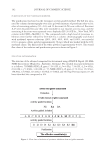
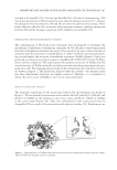
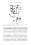
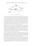
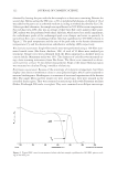
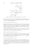
JOURNAL OF COSMETIC SCIENCE 90 causes hyperpigmentation induced by UV irradiation or medical conditions such as melasma, postinfl ammatory hyperpigmentation, and solar lentigo. Therefore, the control of melanogenesis is an important strategy in the treatment of abnormal skin pigmenta- tion for cosmetic and medical purposes (2). Most controllers of melanogenesis act not only by altering tyrosinase gene expression but also by altering the mechanisms responsible for the transfer of melanosomes to keratino- cytes (3). The melanocyte-specifi c enzymes, tyrosinase and tyrosinase-related protein 1 and 2 (TRP-1 and TRP-2), are involved in converting tyrosine into melanin pigments (4,5). In particular, tyrosinase catalyzes two rate-limiting reactions involved in melano- genesis: the hydroxylation of tyrosine to 3,4-dihydroxyphenylalanine (DOPA), and the oxidation of DOPA, which results in the formation to dopaquinone (6). Since tyrosinase is a key enzyme in melanogenesis, it has been the target during the screening of new pigmentation inhibitors from natural products. Some examples of these products include kojic acid, arbutin, ascorbic acid derivatives, retinoic acid, azelaic acid, hydroquinone, catechins, aloesin, resveratrol, oxyresveratrol, and 4,4′-dihydroxybiphenyl (7–16). Various plant extracts have recently been studied to fi nd new natural antimelanogenic products. Gastrodia elata (GE), of the Orchidaceae family, is a traditional herbal medicine in East Asian countries. Zhao et al. (17) identifi ed the components of GE Blume using the capillary zone electrophoresis, the fi ve constituents—gastrodin (GA), 4-hydroxybenzyl alcohol (4-HBA), vanillyl alcohol, 4-hydroxybenzaldehyde (HD), and vanillin—in the extracts of GE Blume roots. It has been used for centuries as an anticonvulsant, analgesic, and sedative for the medical treatment of headaches, epilepsy, dizziness, rheumatism, neuralgia, paralysis, hyperten- sion, and other neuralgic disorders (18,19). A previous study showed that the anticonvul- sive effect of GE was attributed to its antioxidant activity, which was due to the antioxidant actions of 4-HBA, one of several phenolic components of GE extract (20). Since GE extract has several phenolic components that have antioxidant activity, this plant has been suggested to also have a depigmenting effect. Our group is currently trying to fi nd new antimelanogenic substances from natural sources. In this regard, GE has long been used in the treatment of many diseases, espe- cially skin diseases, and is well known for its antioxidant effects. In this study, we evaluate the antimelanogenic effects of GE in HM3KO melanoma cells by suppressing the expres- sion of tyrosinase and TRP-1 mRNA and protein levels. MATERIALS AND METHODS GE EXTRACT Plant extracts were purchased from the PURIMED Co., Ltd (Seoul, Korea). The pharma- ceutical name was Gastrodiae Rhizoma, which is the tuberous root extract of the GE Blume. The dried herb recommended in the protocol provided by PURIMED Co. Ltd was purchased from a Chinese market (Hong Kong, China) and used in this study. For the extraction of water-soluble fraction, 200 mg of the plants were cut into small pieces and boiled in 200 ml of distilled water under refl ux conditions. After 1 h, the mixture was fi ltered and the fi ltrate was collected. An additional 250 ml of water was added to the

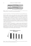
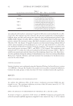
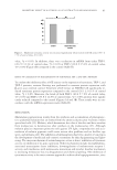
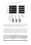
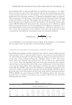
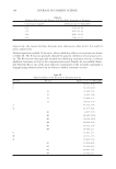
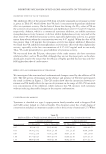
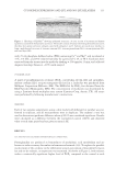
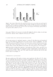
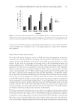
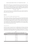
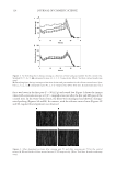
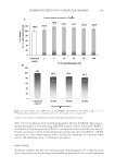
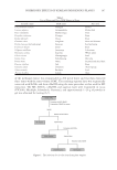
INHIBITORY EFFECT OF GASTRODIA ELATA EXTRACT ON MELANOGENESIS 91 plants residue and boiled for 1 h. The fi ltrate was collected using a 0.2-μm syringe fi lter, combined with the previous fi ltrate sample, and allowed to cool at room temperature. The extracts were stored in the matrix tube and then sterilized using an autoclave. TYROSINASE INHIBITION ASSAY Tyrosinase activity was measured by its DOPA oxidase activity. The tyrosinase inhibition in a cell-free system was tested by mushroom tyrosinase (Sigma, St. Louis, MO). Preincu- bation was conducted with 0.05 ml of 2000 U/ml of mushroom tyrosinase in a 0.1 M phosphate buffer at pH 6.5, followed by incubation in 0.05 M phosphate buffer with the varied concentrations of GE extracts (100, 500, and 1000 μg/ml) and 3 mM L -tyrosine for 10 min at 37°C. The tyrosinase activity was determined by optical density measured at 475 nm using an ELISA microplate reader (Molecular Device Ltd, Wokingham, United Kingdom) and is expressed as a percentage of the control value (100%) an arbutin was used as the reference. CELL CULTURE A pigmented melanoma cell line, HM3KO, established by Ohashi et al. (21) from meta- static melanoma cells of peritoneal fl uids was kindly supplied by Yoko Funasaka (Kobe University School of Medicine, Kobe, Japan). Cells were grown in culture medium con- sisting of Dulbecco’s modifi ed Eagle’s medium with 10% fetal bovine serum (Gibco, Grand Island, NY) and 100 U/ml penicillin–streptomycin in a humidifi ed incubator with 5% CO2 at 37°C. The cells were regularly observed using an inverted microscope and seeded onto 60-mm dishes at a density of 1 × 10⁵ cells per dish. RNA EXTRACTION AND REVERSE TRANSCRIPTION POLYMERASE CHAIN REACTION (RT-PCR) Total RNA was extracted from HM3KO melanoma cells using TRIzol Reagent accord- ing to the manufacturer’s protocol. The RNA samples (2 μg per reaction) were reverse transcribed with reverse transcriptase (Promega, Madison, WI) in the presence of oligo- dT, and the RT product was used for amplifi cation with Taq polymerase. The resulting cDNA was amplifi ed using specifi c primers (Bioneer, Daejeon, Korea) (Table I). Specifi c primers for glyceraldehyde-3-phosphate dehydrogenase (GAPDH) were added as an in- ternal standard for the same reverse transcriptase product. Amplifi cation conditions were 94°C (30 s), 60°C (30 s), and 72°C (40 s) for 35 cycles (tyrosinase), 25 cycles (TRP-1, TRP-2), and 20 cycles (GAPDH), respectively. The PCR products were separated by electrophoresis on 1.2% agarose gels and stained with ethidium bromide. WESTERN BLOT ANALYSIS The HM3KO melanoma cells were seeded onto 60-mm dishes at a density of 1 × 10⁵ cells per dish and cultivated by the method described above. After incubation for 24 h, the medium
Purchased for the exclusive use of nofirst nolast (unknown)
From: SCC Media Library & Resource Center (library.scconline.org)